Copyright
©The Author(s) 2020.
World J Clin Cases. Oct 26, 2020; 8(20): 4773-4784
Published online Oct 26, 2020. doi: 10.12998/wjcc.v8.i20.4773
Published online Oct 26, 2020. doi: 10.12998/wjcc.v8.i20.4773
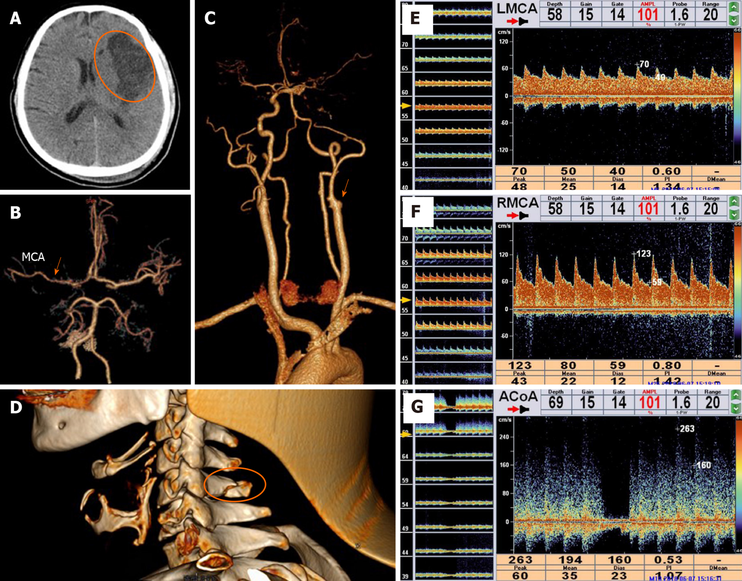
Figure 4 Imaging of case 3.
A: Computed tomography (CT) revealed left frontotemporal infarction (ellipse); B: Head CT angiography revealed a thin middle cerebral artery (arrow); C: Cervical CT angiography revealed left internal carotid artery occlusion (arrow); D: A 3D-CT scan showed a cervical spinous process fracture (ellipse); E and F: Transcranial doppler showed asymmetry between the blood flow velocities of the two middle cerebral artery caused by left traumatic internal carotid artery dissection; G: When the right internal carotid artery was subjected to a neck compression test, the velocity of the anterior communicating artery rapidly decreased to zero and recovered when the compression was released. In E-G: FVd is the diastolic blood flow velocity, FVm is the mean blood flow velocity, FVs is the systolic blood flow velocity, and PI is the pulsatility index. MCA: Middle cerebral artery.
- Citation: Wang GM, Xue H, Guo ZJ, Yu JL. Cerebral infarct secondary to traumatic internal carotid artery dissection. World J Clin Cases 2020; 8(20): 4773-4784
- URL: https://www.wjgnet.com/2307-8960/full/v8/i20/4773.htm
- DOI: https://dx.doi.org/10.12998/wjcc.v8.i20.4773


