Copyright
©The Author(s) 2020.
World J Clin Cases. Oct 26, 2020; 8(20): 4763-4772
Published online Oct 26, 2020. doi: 10.12998/wjcc.v8.i20.4763
Published online Oct 26, 2020. doi: 10.12998/wjcc.v8.i20.4763
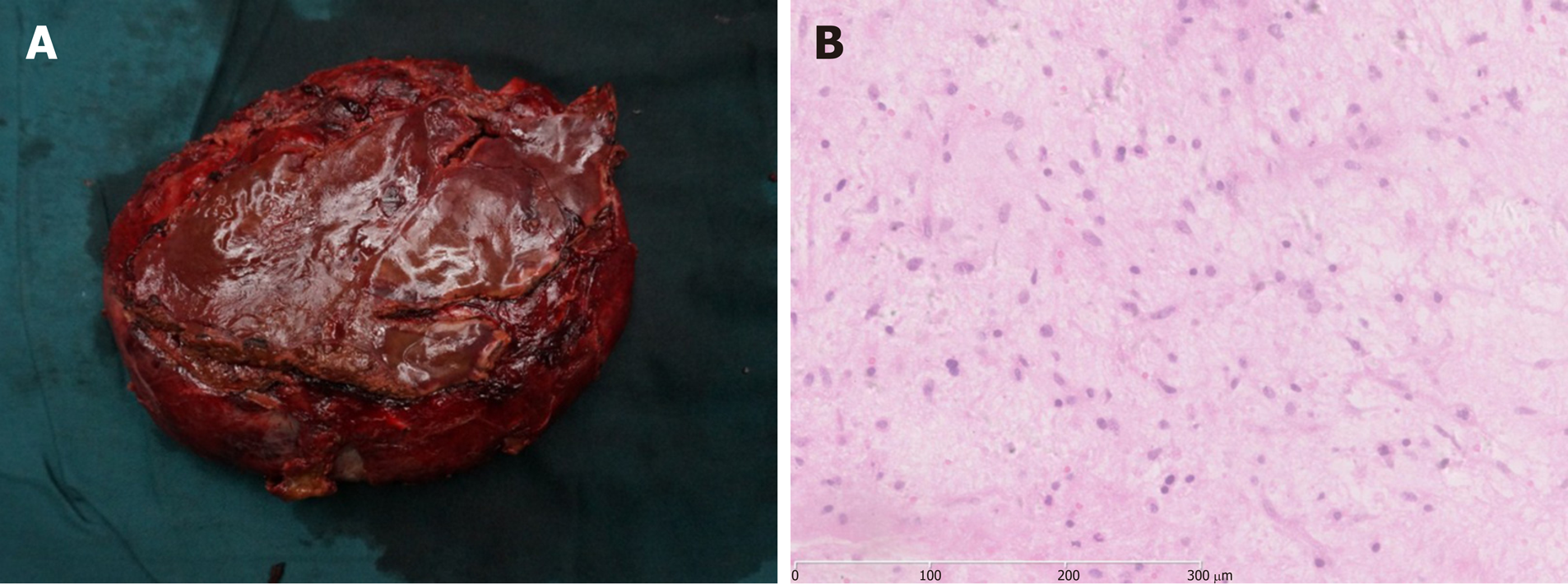
Figure 2 Pathological findings of undifferentiated embryonal liver sarcoma.
A: Macroscopically, the large cystic mass with an amount of dark red blood clots in the lumen was observed; B: Microscopically, the tumor was composed of pleomorphic and polynuclear dyskaryotic cells, and mesenchymal hamartomalike lesions were partially seen (hematoxylin and eosin staining).
- Citation: Zhang C, Jia CJ, Xu C, Sheng QJ, Dou XG, Ding Y. Undifferentiated embryonal sarcoma of the liver: Clinical characteristics and outcomes. World J Clin Cases 2020; 8(20): 4763-4772
- URL: https://www.wjgnet.com/2307-8960/full/v8/i20/4763.htm
- DOI: https://dx.doi.org/10.12998/wjcc.v8.i20.4763


