Copyright
©The Author(s) 2020.
World J Clin Cases. Oct 26, 2020; 8(20): 4763-4772
Published online Oct 26, 2020. doi: 10.12998/wjcc.v8.i20.4763
Published online Oct 26, 2020. doi: 10.12998/wjcc.v8.i20.4763
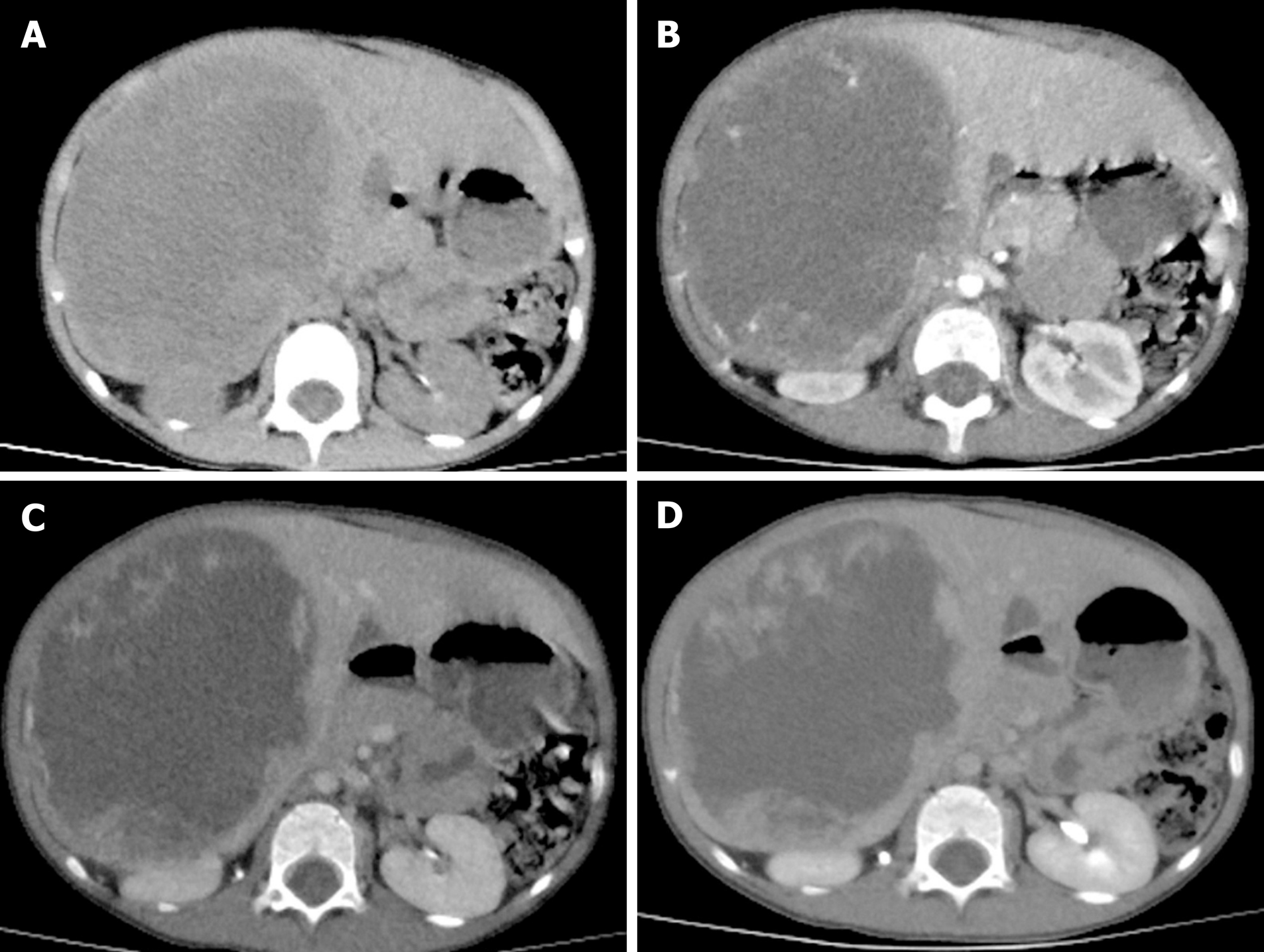
Figure 1 Computed tomography images of undifferentiated embryonal liver sarcoma.
A: Plain CT scan revealed hepatomegaly. In the right lobe of the liver, there was a large oval cystic-solid mixed mass measruing 12.0 cm × 8.8 cm × 12.2 cm, with a clear boundary and non-homogeneous density; the solid part had multiple nodular soft tissue density shadows; B: In the arterial phase, the cystic components of the mass were not obviously strengthened; however, the solid components of the mass were obviously strengthened; C: In the portal venous phase, the enhanced extent of the masses was a little more obvious than that in the arterial phase; The enhancement of the masses was significantly lower than that of surrounding parenchyma; D: In the delayed phase, the enhanced extent of the masses was still a little more obvious than that in the arterial phase.
- Citation: Zhang C, Jia CJ, Xu C, Sheng QJ, Dou XG, Ding Y. Undifferentiated embryonal sarcoma of the liver: Clinical characteristics and outcomes. World J Clin Cases 2020; 8(20): 4763-4772
- URL: https://www.wjgnet.com/2307-8960/full/v8/i20/4763.htm
- DOI: https://dx.doi.org/10.12998/wjcc.v8.i20.4763


