Copyright
©The Author(s) 2020.
World J Clin Cases. Oct 26, 2020; 8(20): 4708-4718
Published online Oct 26, 2020. doi: 10.12998/wjcc.v8.i20.4708
Published online Oct 26, 2020. doi: 10.12998/wjcc.v8.i20.4708
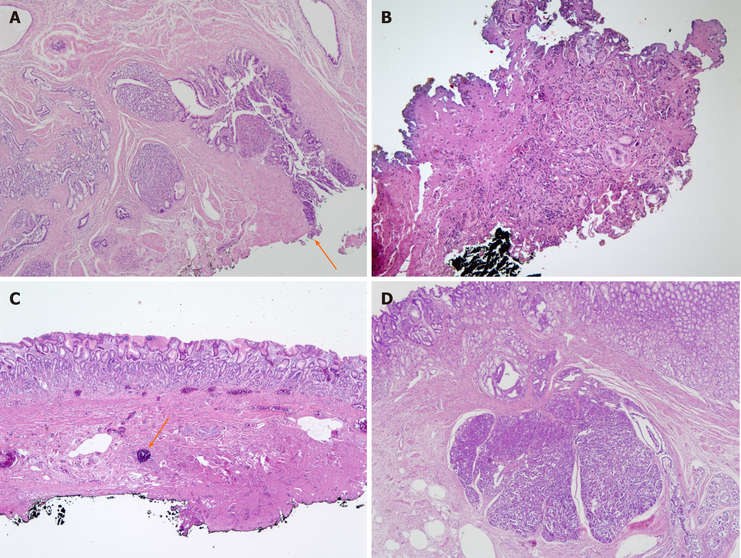
Figure 4 Representative histologic images of gastric heterotopic pancreas.
A: Patient 1: Pancreatic tissue is in proper muscle with involvement of resection margin (arrow) (hematoxylin-eosin; original magnification, × 40); B: Patient 3: There is focal nest of cells and bluish material with fibrosis and severe cautery artifact (× 100); C: Patient 4: Submucosal fibrosis with foreign body reaction and dystrophic calcification (arrow) was noted (× 40); D: Patient 5: Pancreatic tissue is in submucosa overlying gastric mucosa (× 40).
- Citation: Noh JH, Kim DH, Kim SW, Park YS, Na HK, Ahn JY, Jung KW, Lee JH, Choi KD, Song HJ, Lee GH, Jung HY. Endoscopic submucosal dissection as alternative to surgery for complicated gastric heterotopic pancreas. World J Clin Cases 2020; 8(20): 4708-4718
- URL: https://www.wjgnet.com/2307-8960/full/v8/i20/4708.htm
- DOI: https://dx.doi.org/10.12998/wjcc.v8.i20.4708


