Copyright
©The Author(s) 2020.
World J Clin Cases. Jan 26, 2020; 8(2): 451-463
Published online Jan 26, 2020. doi: 10.12998/wjcc.v8.i2.451
Published online Jan 26, 2020. doi: 10.12998/wjcc.v8.i2.451
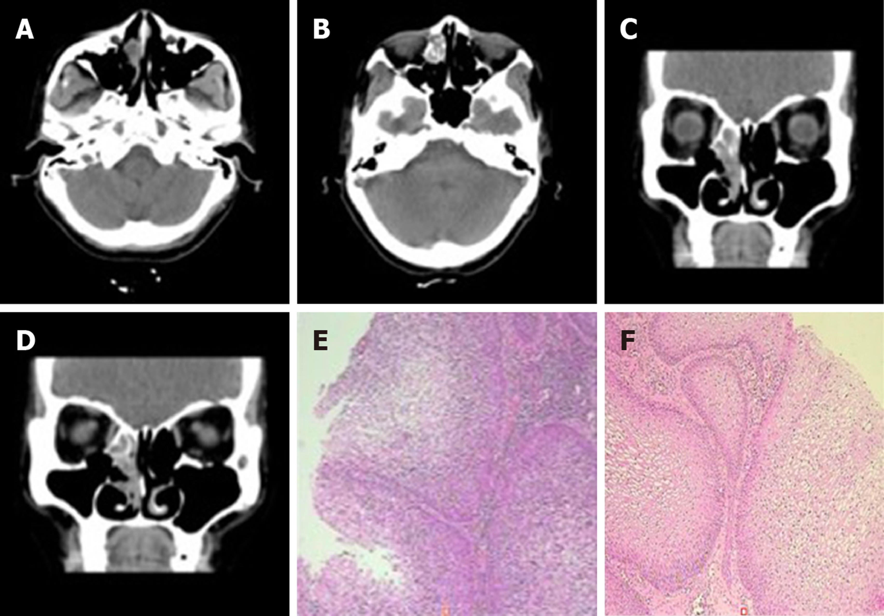
Figure 2 Computed tomography scan and histopathologic examination results of Case 1.
A-D: The first preoperative examination (February 4, 2016). Axial and sagittal paranasal sinus computed tomography scans showed dense shadows in the soft tissue of the right inferior nasal tract with small, low-density shadows, which were indistinct from the inferior turbinate and ethmoid sinus, and bone destruction in the ethmoid sinus; E and F: Histopathologic findings showed that the nasal mass was an inverted papilloma and that the maxillary sinus exhibited chronic inflammation of the mucous membrane.
- Citation: Wang LL, Chen FJ, Yang LS, Li JE. Analysis of pathogenetic process of fungal rhinosinusitis: Report of two cases. World J Clin Cases 2020; 8(2): 451-463
- URL: https://www.wjgnet.com/2307-8960/full/v8/i2/451.htm
- DOI: https://dx.doi.org/10.12998/wjcc.v8.i2.451


