Copyright
©The Author(s) 2020.
World J Clin Cases. Jan 26, 2020; 8(2): 436-443
Published online Jan 26, 2020. doi: 10.12998/wjcc.v8.i2.436
Published online Jan 26, 2020. doi: 10.12998/wjcc.v8.i2.436
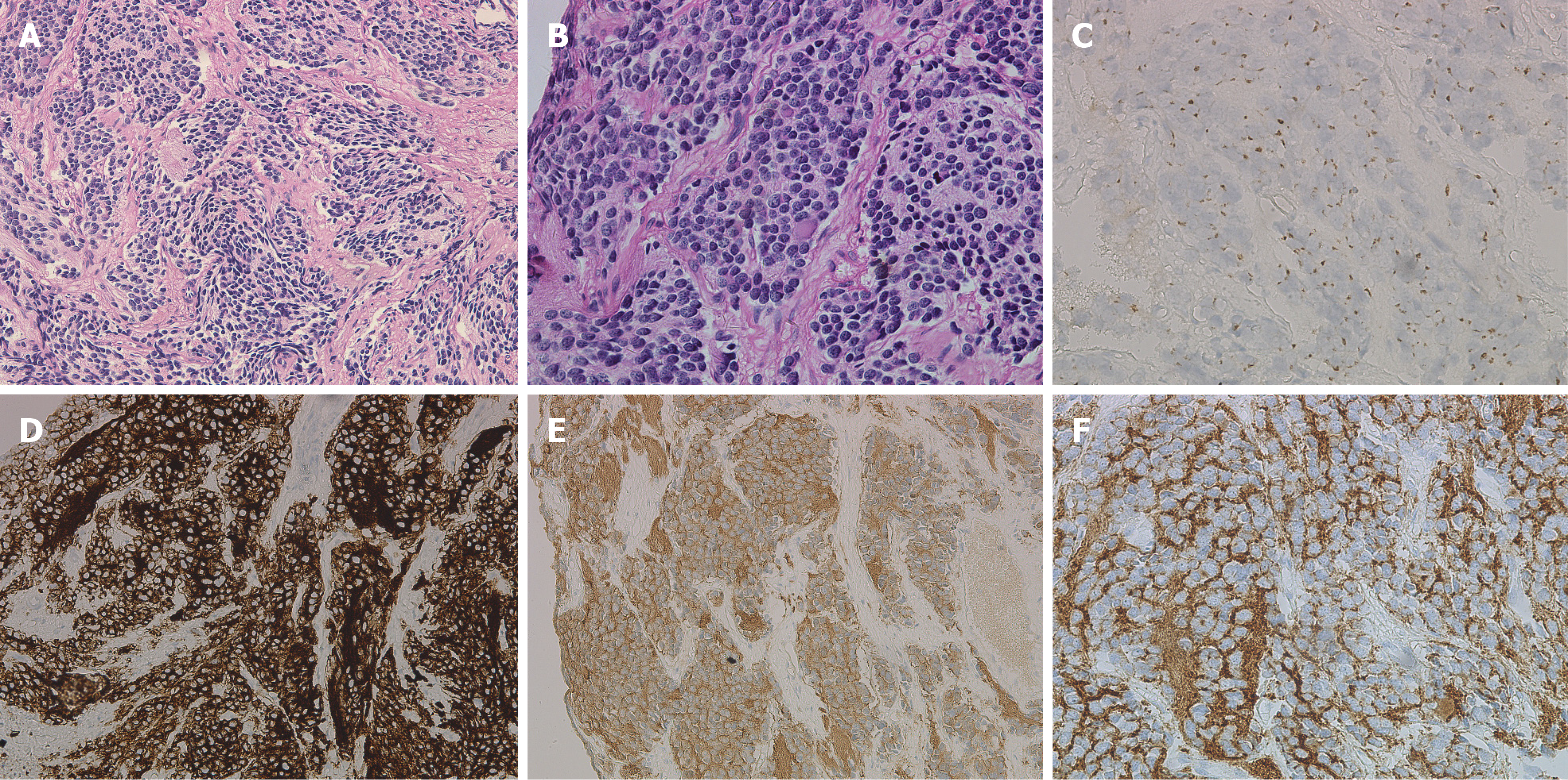
Figure 3 Histological and pathological images of the tumour biopsy specimens.
A: HE staining, ×200; B: HE staining, ×400; C: CgA (suspiciously positive); D: CD56 (positive); E: NSE (positive); F: Syn (positive).
- Citation: Liu J, Wu XW, Hao XW, Duan YH, Wu LL, Zhao J, Zhou XJ, Zhu CZ, Wei B, Dong Q. Spontaneous regression of stage III neuroblastoma: A case report. World J Clin Cases 2020; 8(2): 436-443
- URL: https://www.wjgnet.com/2307-8960/full/v8/i2/436.htm
- DOI: https://dx.doi.org/10.12998/wjcc.v8.i2.436


