Copyright
©The Author(s) 2020.
World J Clin Cases. Jan 26, 2020; 8(2): 425-435
Published online Jan 26, 2020. doi: 10.12998/wjcc.v8.i2.425
Published online Jan 26, 2020. doi: 10.12998/wjcc.v8.i2.425
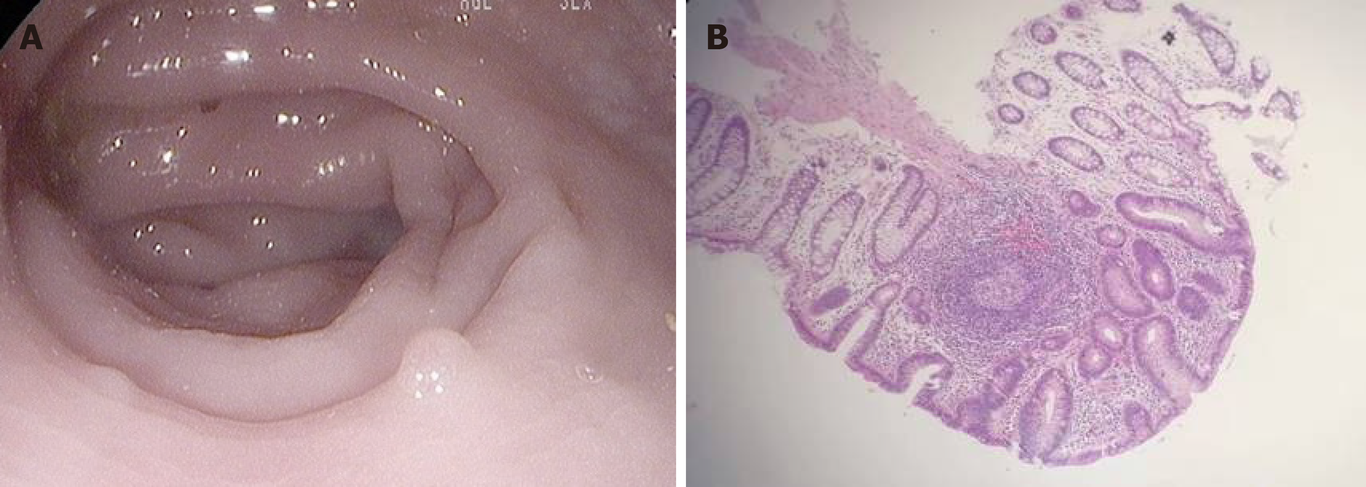
Figure 2 Gut inflammation verified by endoscopic examination.
A: Macroscopic examination of the large intestine showed that the mucous membrane was congested and swollen, the vascular lakes were absent, and there were several polyps in the sigmoid colon and rectum, confirming the presence of gut inflammation; B: Microscopic examination revealed that these polyps were enlarged lymphoid follicles which were structurally integrated, with the infiltration of a large amount of lymphocytes and plasma cells around the lymphoid follicles and underneath the mucous membrane, in accordance with inflammatory proliferation.
- Citation: Zhao XC, Zhao L, Sun XY, Xu ZS, Ju B, Meng FJ, Zhao HG. Excellent response of severe aplastic anemia to treatment of gut inflammation: A case report and review of the literature. World J Clin Cases 2020; 8(2): 425-435
- URL: https://www.wjgnet.com/2307-8960/full/v8/i2/425.htm
- DOI: https://dx.doi.org/10.12998/wjcc.v8.i2.425


