Copyright
©The Author(s) 2020.
World J Clin Cases. Jan 26, 2020; 8(2): 415-424
Published online Jan 26, 2020. doi: 10.12998/wjcc.v8.i2.415
Published online Jan 26, 2020. doi: 10.12998/wjcc.v8.i2.415
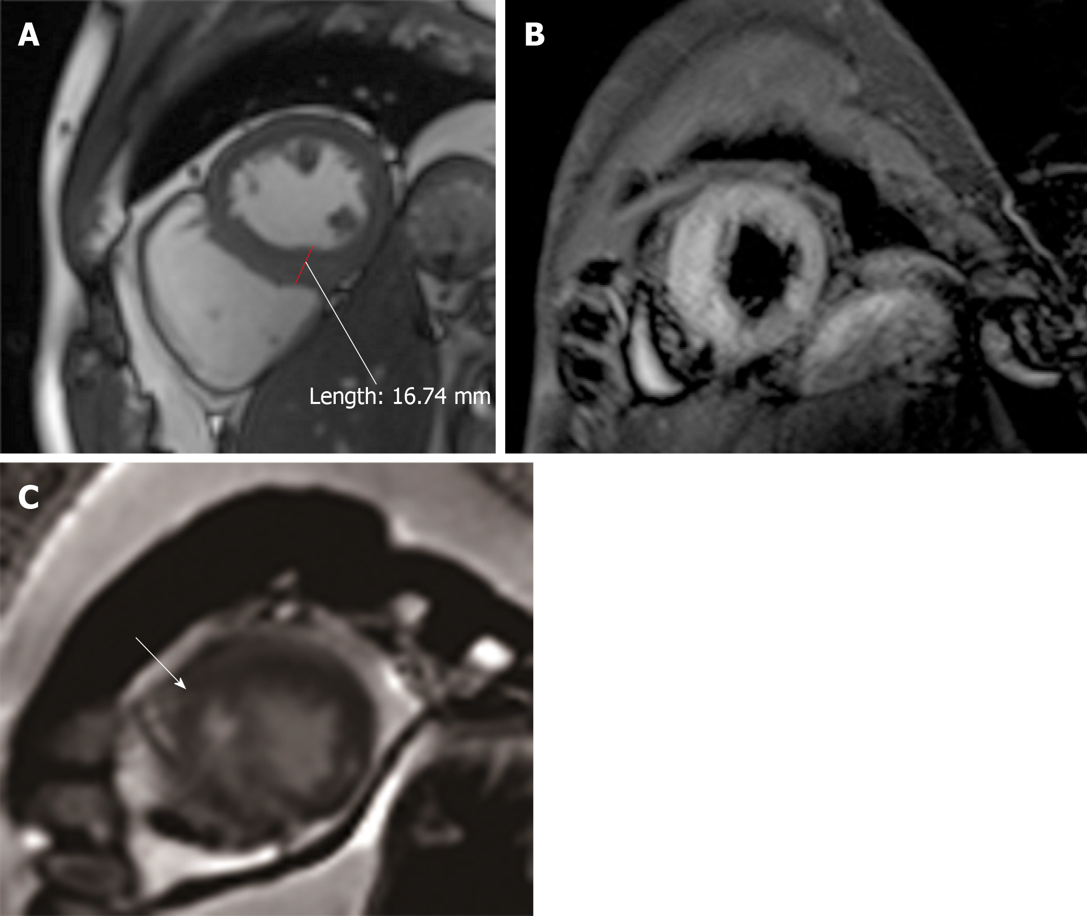
Figure 5 Cardiovascular magnetic resonance imaging.
A: In the CINE sequence at the left ventricular end-diastolic phase, the apical septal wall was less extensive after 12 d; the ventricular wall was 16.74 mm, which was thicker than normal; B: FS-T2WI showed obvious edema; C: The anterior and interior late gadolinium enhancement moved to the middle myocardium.
- Citation: Hou YM, Han PX, Wu X, Lin JR, Zheng F, Lin L, Xu R. Myocarditis presenting as typical acute myocardial infarction: A case report and review of the literature. World J Clin Cases 2020; 8(2): 415-424
- URL: https://www.wjgnet.com/2307-8960/full/v8/i2/415.htm
- DOI: https://dx.doi.org/10.12998/wjcc.v8.i2.415


