Copyright
©The Author(s) 2020.
World J Clin Cases. Jan 26, 2020; 8(2): 382-389
Published online Jan 26, 2020. doi: 10.12998/wjcc.v8.i2.382
Published online Jan 26, 2020. doi: 10.12998/wjcc.v8.i2.382
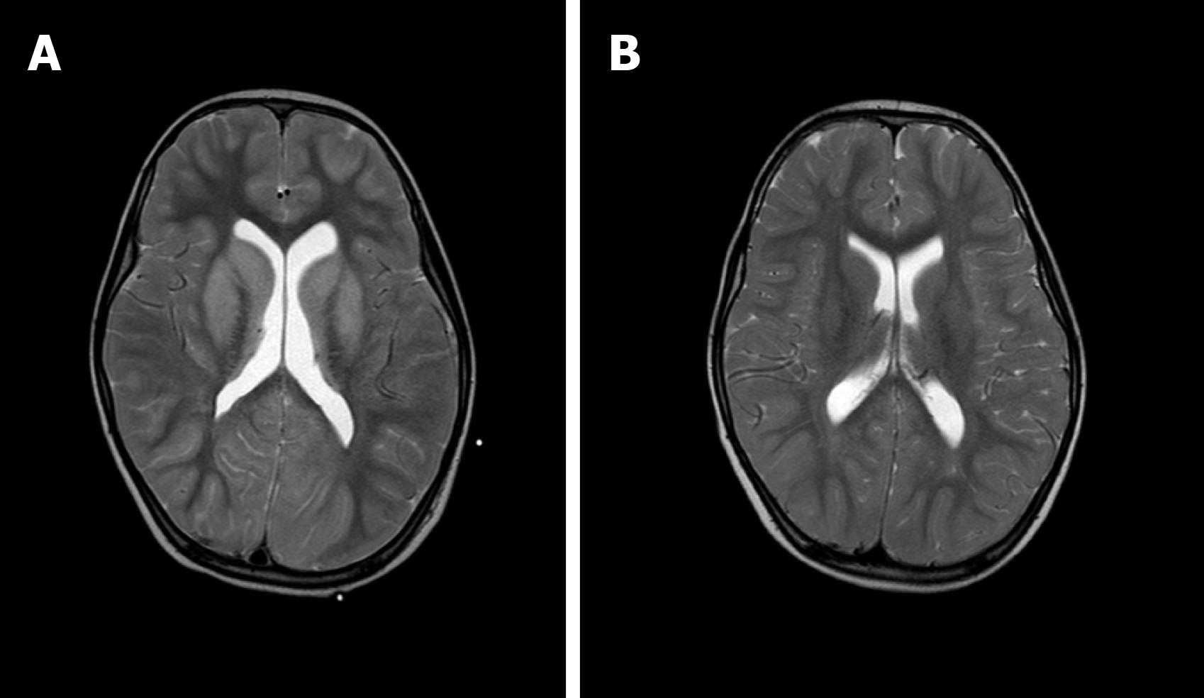
Figure 3 Imaging of the basal ganglia after and before brainstem folding.
A: After brain stem folding, swelling of the bilateral putamen, cauda nucleus, frontal lobe, parietal lobe, and occipital cortex, increased T2WI signals, and slightly enlarged bilateral lateral ventricle can be seen; B: Three days before brain stem folding, no swelling was observed in the bilateral putamen, caudate nucleus, frontal lobe, parietal lobe, or occipital cortex, with normal signals, and no expansion was observed in the bilateral lateral ventricles.
- Citation: Li SY, Li PQ, Xiao WQ, Liu HS, Yang SD. Brainstem folding in an influenza child with Dandy-Walker variant. World J Clin Cases 2020; 8(2): 382-389
- URL: https://www.wjgnet.com/2307-8960/full/v8/i2/382.htm
- DOI: https://dx.doi.org/10.12998/wjcc.v8.i2.382


