Copyright
©The Author(s) 2020.
World J Clin Cases. Jan 26, 2020; 8(2): 353-361
Published online Jan 26, 2020. doi: 10.12998/wjcc.v8.i2.353
Published online Jan 26, 2020. doi: 10.12998/wjcc.v8.i2.353
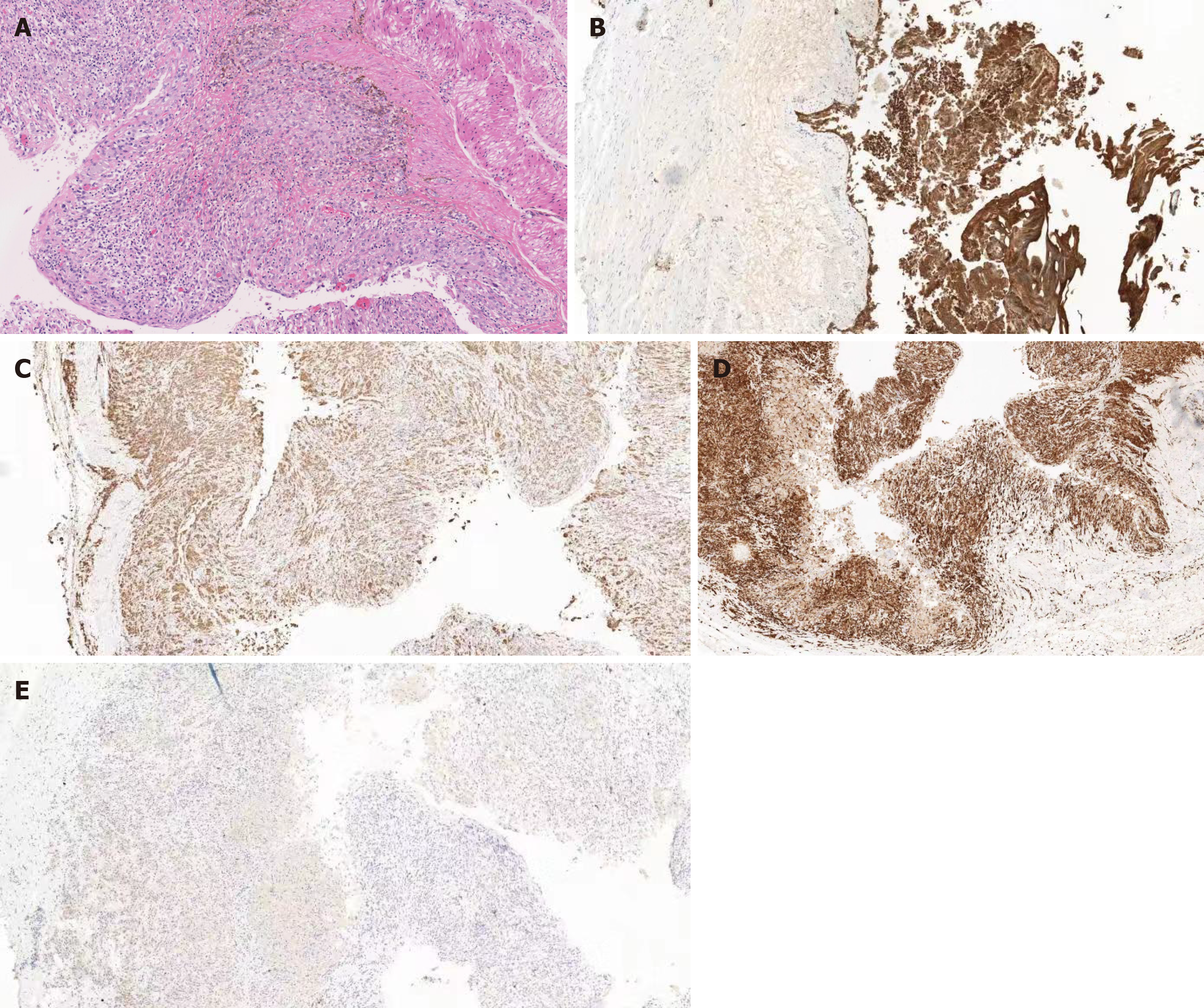
Figure 3 Histological examinations showed the specimen was consistent with bronchogenic cyst with obvious hyperplasia of histiocytes, and no dysplasia/malignancy was found.
A: HE stains × 200. B: CK (pan) (epithelium +) × 200; C: CD68 (histocyte +) × 200; D: CD163 (histocyte +) × 200; E: S-100 (-) × 200.
- Citation: Zhang FM, Chen HT, Ning LG, Xu Y, Xu GQ. Esophageal bronchogenic cyst excised by endoscopic submucosal tunnel dissection: A case report. World J Clin Cases 2020; 8(2): 353-361
- URL: https://www.wjgnet.com/2307-8960/full/v8/i2/353.htm
- DOI: https://dx.doi.org/10.12998/wjcc.v8.i2.353


