Copyright
©The Author(s) 2020.
World J Clin Cases. Jan 26, 2020; 8(2): 353-361
Published online Jan 26, 2020. doi: 10.12998/wjcc.v8.i2.353
Published online Jan 26, 2020. doi: 10.12998/wjcc.v8.i2.353
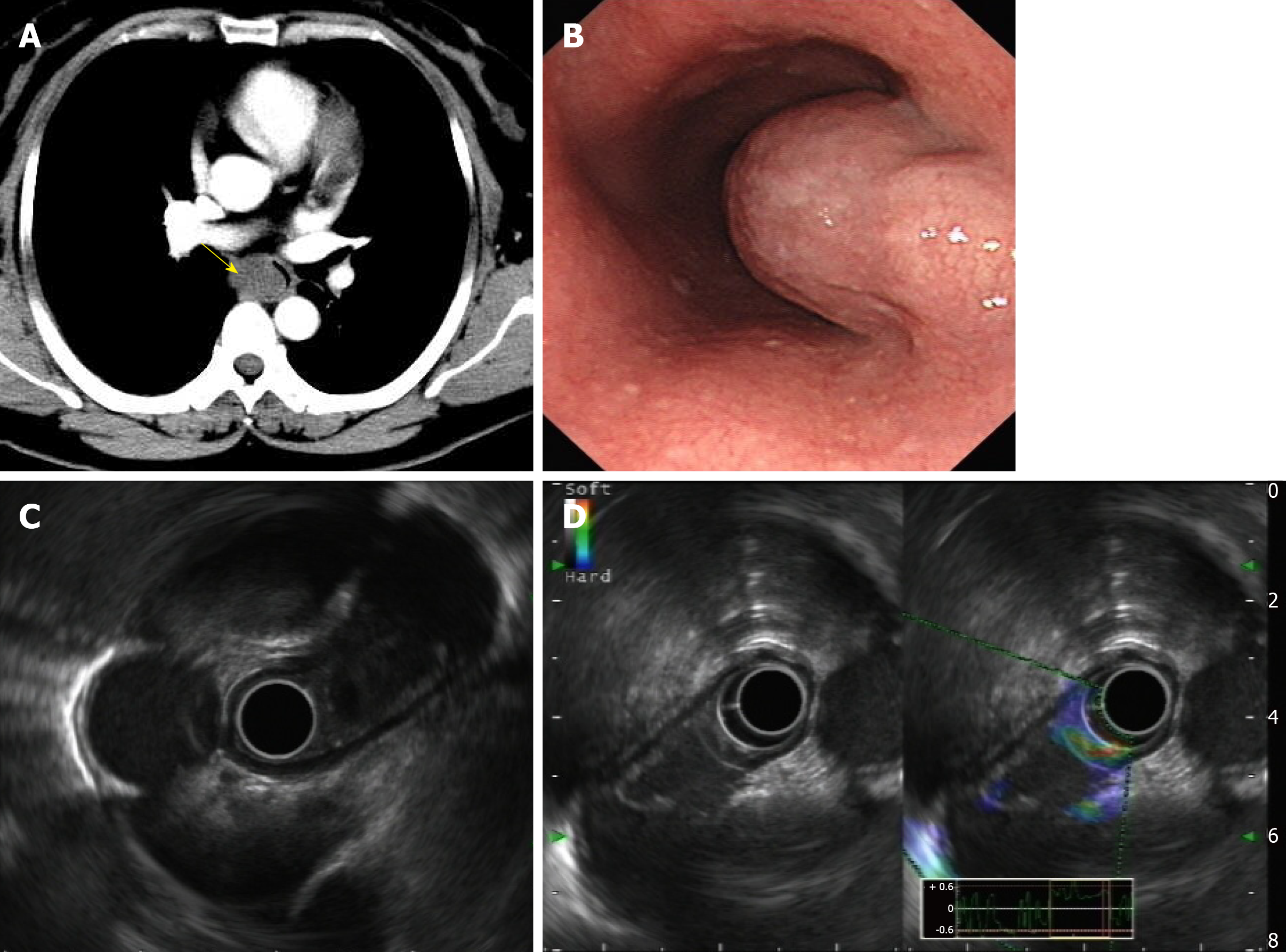
Figure 1 Enhanced thoracic computer tomography and endoscopic ultrasonography.
A: A slightly oval-shaped low-density lesion with clear boundary in the upper middle part of the esophagus in the enhanced thoracic computer tomography (yellow arrow); B: A submucosal mass was observed about 28 cm from the incisor with a gourd-like appearance; C: Probe EUS view; D: Contrast-enhanced ultrasonography showed slight enhancement around the lesion but no internal enhancement.
- Citation: Zhang FM, Chen HT, Ning LG, Xu Y, Xu GQ. Esophageal bronchogenic cyst excised by endoscopic submucosal tunnel dissection: A case report. World J Clin Cases 2020; 8(2): 353-361
- URL: https://www.wjgnet.com/2307-8960/full/v8/i2/353.htm
- DOI: https://dx.doi.org/10.12998/wjcc.v8.i2.353


