Copyright
©The Author(s) 2020.
World J Clin Cases. Jan 26, 2020; 8(2): 325-330
Published online Jan 26, 2020. doi: 10.12998/wjcc.v8.i2.325
Published online Jan 26, 2020. doi: 10.12998/wjcc.v8.i2.325
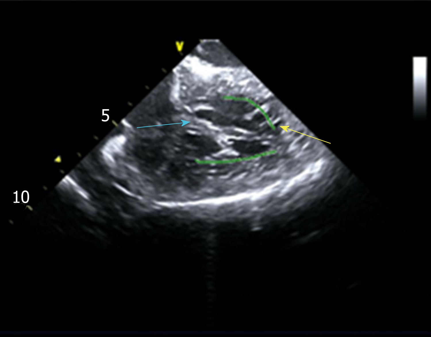
Figure 2 Left ventricular false tendons were detected by intracardiac two-dimensional echocardiography.
The yellow arrow indicates the ventricular apex attachment spot of the left ventricular false tendon, and the biue arrow indicates the basal side of the interventricular septum attachment spot of the left ventricular false tendon.
- Citation: Yang YB, Li XF, Guo TT, Jia YH, Liu J, Tang M, Fang PH, Zhang S. Catheter ablation of premature ventricular complexes associated with false tendons: A case report. World J Clin Cases 2020; 8(2): 325-330
- URL: https://www.wjgnet.com/2307-8960/full/v8/i2/325.htm
- DOI: https://dx.doi.org/10.12998/wjcc.v8.i2.325


