Copyright
©The Author(s) 2020.
World J Clin Cases. Oct 6, 2020; 8(19): 4667-4675
Published online Oct 6, 2020. doi: 10.12998/wjcc.v8.i19.4667
Published online Oct 6, 2020. doi: 10.12998/wjcc.v8.i19.4667
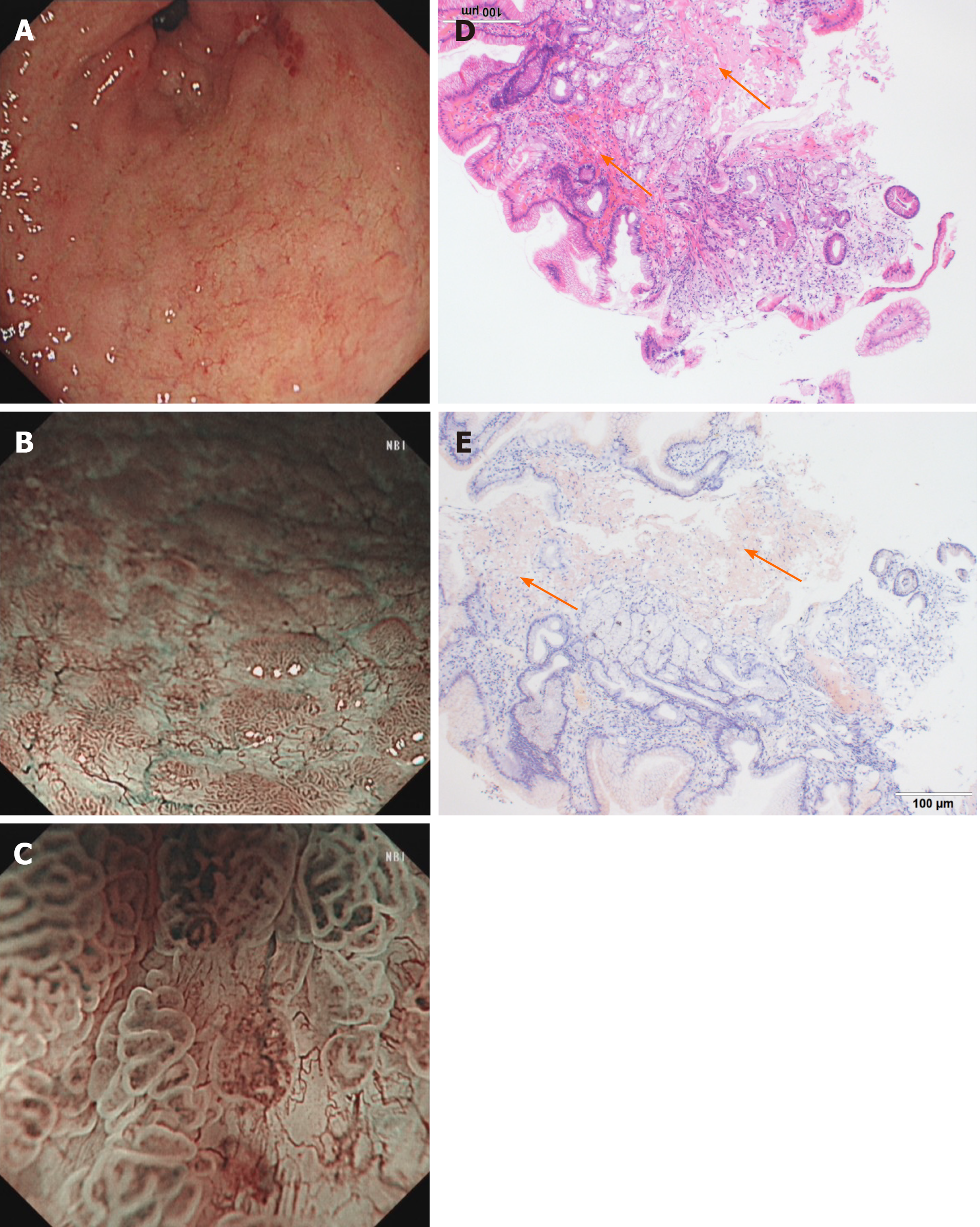
Figure 2 Endoscopic and histological data of patient 2.
A: Conventional endoscopy showed a red and white area in the atrophic gastric antrum with active gastritis. B: Narrow-band imaging (NBI) showed a defined light brownish area. C: Magnifying endoscopy with NBI revealed expanded normal glands with changed polarity and tree-like vessels. D: Hematoxylin-eosin staining showed abundant cord-like red substances in the mucosal layers. E: These tissues were positive for Congo red staining. Orange arrows indicate amyloid depositions.
- Citation: Liu XM, Di LJ, Zhu JX, Wu XL, Li HP, Wu HC, Tuo BG. Localized primary gastric amyloidosis: Three case reports. World J Clin Cases 2020; 8(19): 4667-4675
- URL: https://www.wjgnet.com/2307-8960/full/v8/i19/4667.htm
- DOI: https://dx.doi.org/10.12998/wjcc.v8.i19.4667


