Copyright
©The Author(s) 2020.
World J Clin Cases. Oct 6, 2020; 8(19): 4652-4659
Published online Oct 6, 2020. doi: 10.12998/wjcc.v8.i19.4652
Published online Oct 6, 2020. doi: 10.12998/wjcc.v8.i19.4652
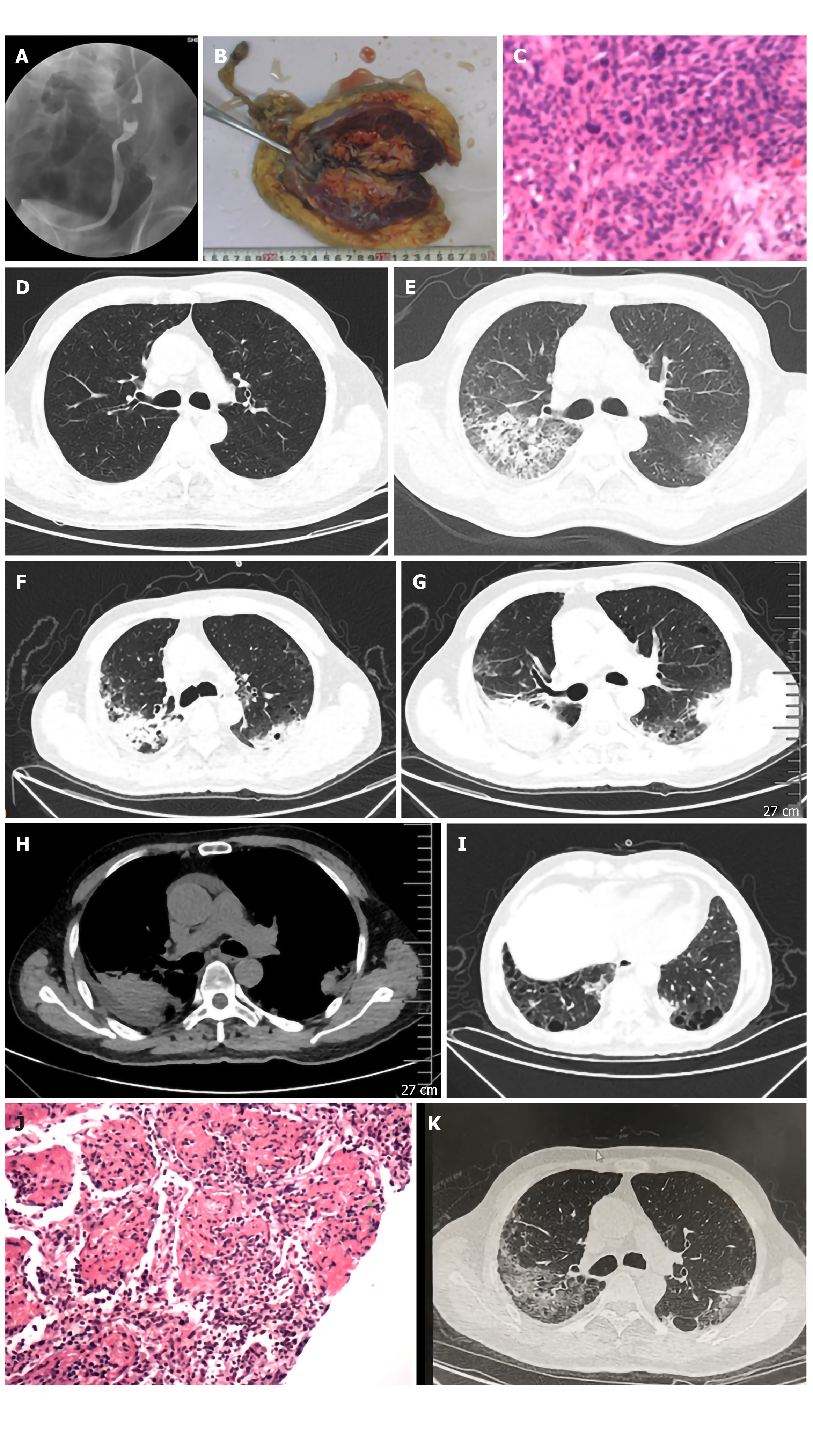
Figure 1 Radiologic and pathological examinations of the patient with intravesical gemcitabine-induced pulmonary toxicity.
A: Ureterography indicating left urothelial obstruction; B: Gross appearance of urothelial carcinoma after laparoscopic radical nephroureterectomy with bladder cuff excision; C: Pathological examination by hematoxylin and eosin staining showing invasive urothelial carcinoma with infiltration to the muscular layer (× 100); D: Chest computed tomography (CT) scan showing bilateral emphysema and bullae 3 mo before the onset of gemcitabine-induced pulmonary toxicity; E: Chest CT scan on admission showing bilateral patchy infiltration accompanied by preexisting emphysema and bullae; F-I: Chest CT scan on the 11th day showing worsening of the patchy infiltration with foci that were more consolidated after empirical antibiotic therapy, resulting in worsening of the respiratory distress; J: Pathological examination by hematoxylin and eosin staining showing organizing pneumonitis in the percutaneous lung biopsy of the consolidation focus (× 100); K: Chest CT scan 1 mo after admission showing obvious absorption of the multiple consolidation foci after tapered corticosteroid therapy.
- Citation: Zhou XM, Wu C, Gu X. Intravesically instilled gemcitabine-induced lung injury in a patient with invasive urothelial carcinoma: A case report. World J Clin Cases 2020; 8(19): 4652-4659
- URL: https://www.wjgnet.com/2307-8960/full/v8/i19/4652.htm
- DOI: https://dx.doi.org/10.12998/wjcc.v8.i19.4652


