Copyright
©The Author(s) 2020.
World J Clin Cases. Oct 6, 2020; 8(19): 4644-4651
Published online Oct 6, 2020. doi: 10.12998/wjcc.v8.i19.4644
Published online Oct 6, 2020. doi: 10.12998/wjcc.v8.i19.4644
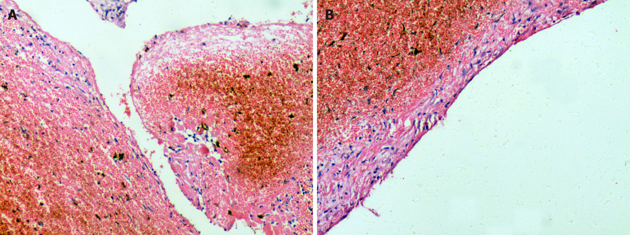
Figure 3 Microscopic examination with haematoxylin and eosin staining was performed.
A: No signs of epithelium were found, although a fibrous tissue lining was observed; B: Remote hemorrhage and clot organization supported the diagnosis of hemophilic pseudotumor (magnification, × 100).
- Citation: Cai X, Yu JJ, Tian H, Shan ZF, Liu XY, Jia J. Intraosseous venous malformation of the maxilla after enucleation of a hemophilic pseudotumor: A case report. World J Clin Cases 2020; 8(19): 4644-4651
- URL: https://www.wjgnet.com/2307-8960/full/v8/i19/4644.htm
- DOI: https://dx.doi.org/10.12998/wjcc.v8.i19.4644


