Copyright
©The Author(s) 2020.
World J Clin Cases. Oct 6, 2020; 8(19): 4644-4651
Published online Oct 6, 2020. doi: 10.12998/wjcc.v8.i19.4644
Published online Oct 6, 2020. doi: 10.12998/wjcc.v8.i19.4644
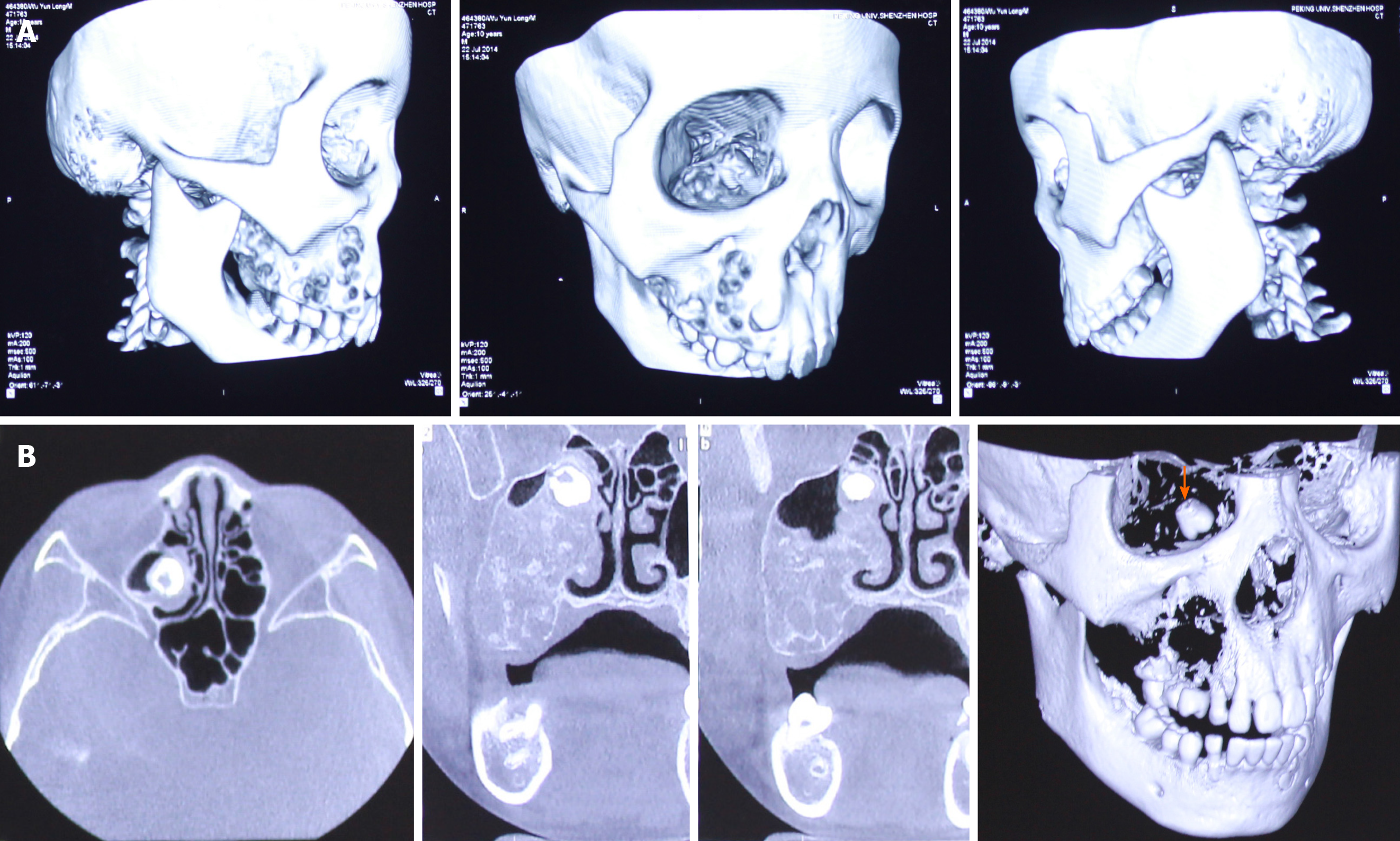
Figure 2 Computed tomography scan and 3D reconstruction displayed a multicystic low-density shadow in the right maxilla.
A: The body of the right maxilla manifested irregular swelling; B: The orange arrow shows a molar that was squeezed into the right maxillary sinus and embedded in the superior part of the lesion close to the canalis opticus.
- Citation: Cai X, Yu JJ, Tian H, Shan ZF, Liu XY, Jia J. Intraosseous venous malformation of the maxilla after enucleation of a hemophilic pseudotumor: A case report. World J Clin Cases 2020; 8(19): 4644-4651
- URL: https://www.wjgnet.com/2307-8960/full/v8/i19/4644.htm
- DOI: https://dx.doi.org/10.12998/wjcc.v8.i19.4644


