Copyright
©The Author(s) 2020.
World J Clin Cases. Oct 6, 2020; 8(19): 4615-4623
Published online Oct 6, 2020. doi: 10.12998/wjcc.v8.i19.4615
Published online Oct 6, 2020. doi: 10.12998/wjcc.v8.i19.4615
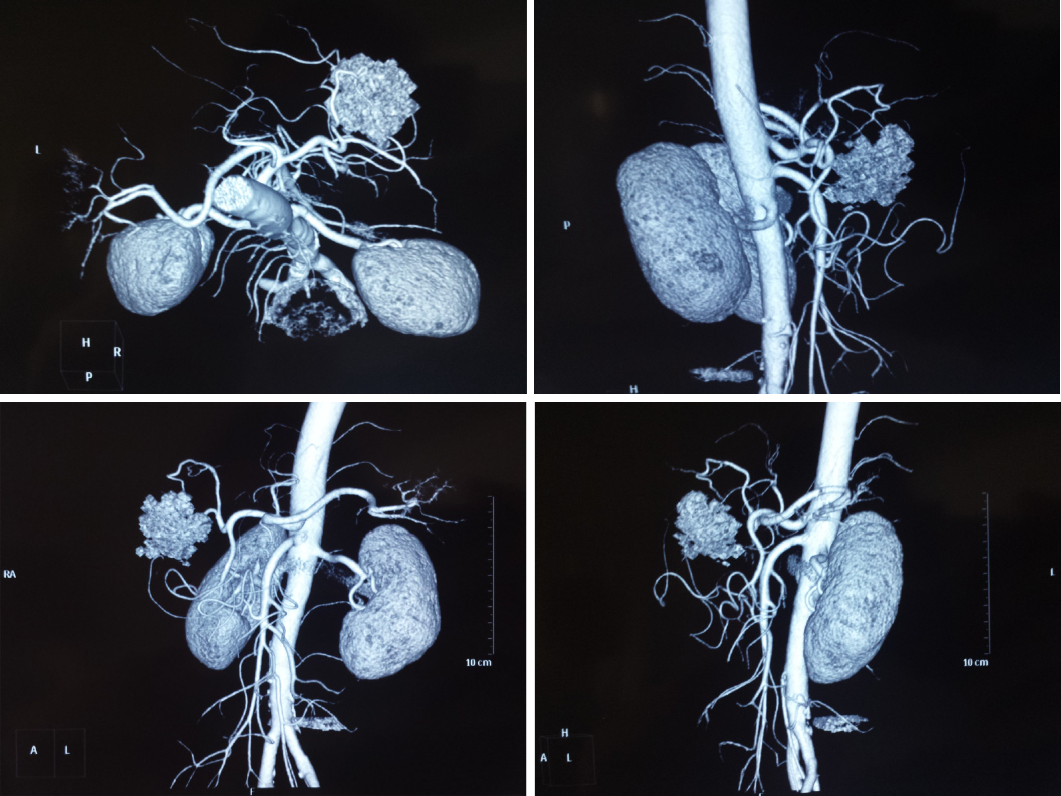
Figure 2 Imaging of hepatic myelolipoma by computed tomography angiography.
Angiography imaging presented a mass in the liver with a rich blood supply that did not correlate to the main branches of the hepatic arteries.
- Citation: Li KY, Wei AL, Li A. Primary hepatic myelolipoma: A case report and review of the literature. World J Clin Cases 2020; 8(19): 4615-4623
- URL: https://www.wjgnet.com/2307-8960/full/v8/i19/4615.htm
- DOI: https://dx.doi.org/10.12998/wjcc.v8.i19.4615


