Copyright
©The Author(s) 2020.
World J Clin Cases. Oct 6, 2020; 8(19): 4588-4594
Published online Oct 6, 2020. doi: 10.12998/wjcc.v8.i19.4588
Published online Oct 6, 2020. doi: 10.12998/wjcc.v8.i19.4588
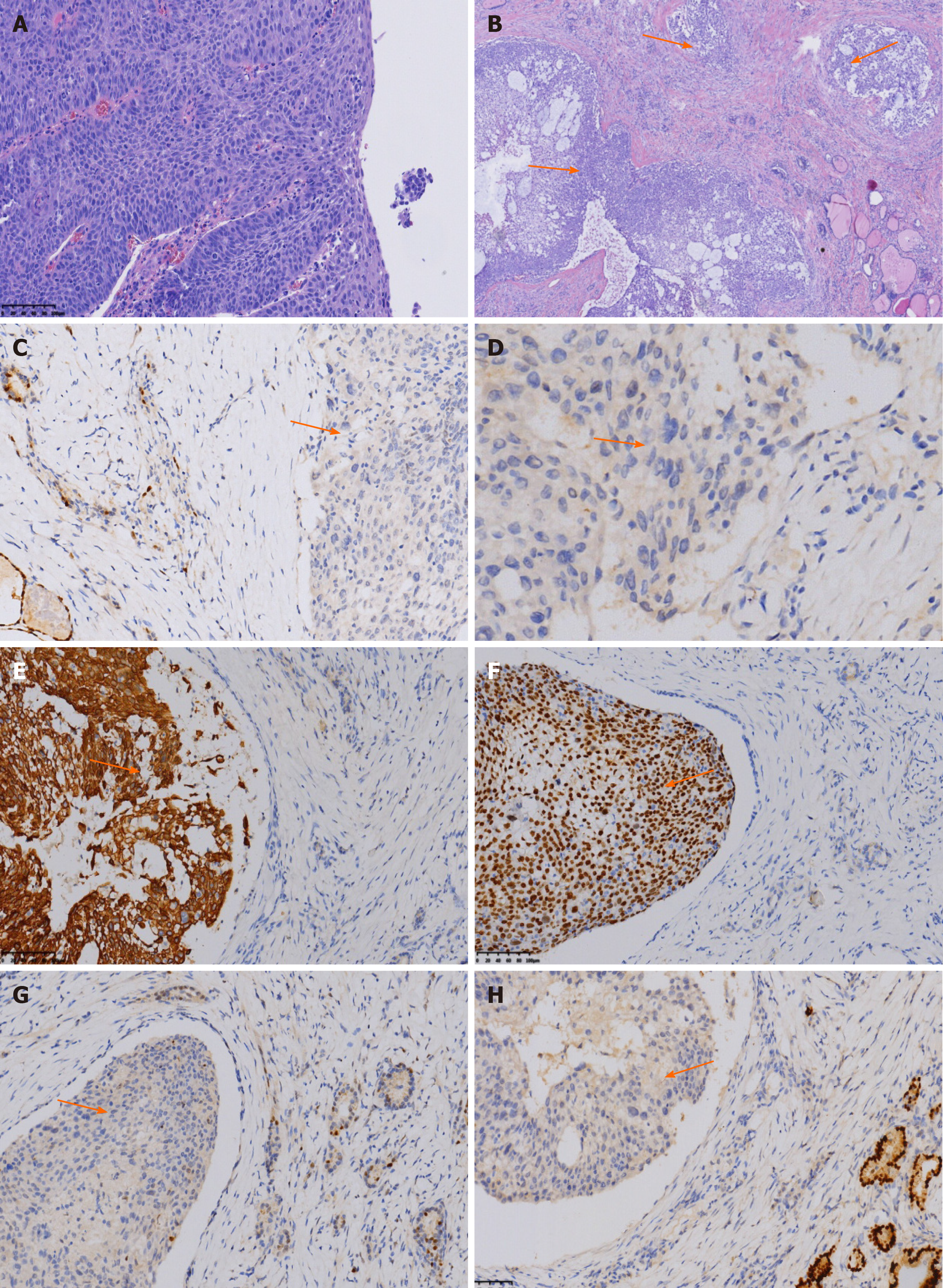
Figure 5 Fine needle aspiration cytology of the thyroid lesions stained with hematoxylin and eosin (× 200) and the immunohistochemical features.
A: Permanent section of the esophagus stained with hematoxylin and eosin shows esophageal squamous cell carcinoma (× 200); B: Histology of MTG stained with hematoxylin and eosin shows multifocal tumor nests (orange arrow) (× 40); C: Immunohistochemical negative staining of metastasis to the thyroid gland (MTG) for thyroid transcription factor-1 (orange arrow) (× 200); D: Immunohistochemical negative staining of MTG for thyroglobulin (orange arrow) (× 200); E: Immunohistochemical positive staining of MTG for CK5/6 (orange arrow) (× 200); F: Immunohistochemical positive staining of MTG for P40 (orange arrow) (× 200); G: Immunohistochemical negative staining of MTG for (orange arrow) (× 200); H: Immunohistochemical negative staining of MTG for paired box gene 8 (orange arrow) (× 200).
- Citation: Zhang X, Gu X, Li JG, Hu XJ. Metastasis of esophageal squamous cell carcinoma to the thyroid gland with widespread nodal involvement: A case report. World J Clin Cases 2020; 8(19): 4588-4594
- URL: https://www.wjgnet.com/2307-8960/full/v8/i19/4588.htm
- DOI: https://dx.doi.org/10.12998/wjcc.v8.i19.4588


