Copyright
©The Author(s) 2020.
World J Clin Cases. Oct 6, 2020; 8(19): 4579-4587
Published online Oct 6, 2020. doi: 10.12998/wjcc.v8.i19.4579
Published online Oct 6, 2020. doi: 10.12998/wjcc.v8.i19.4579
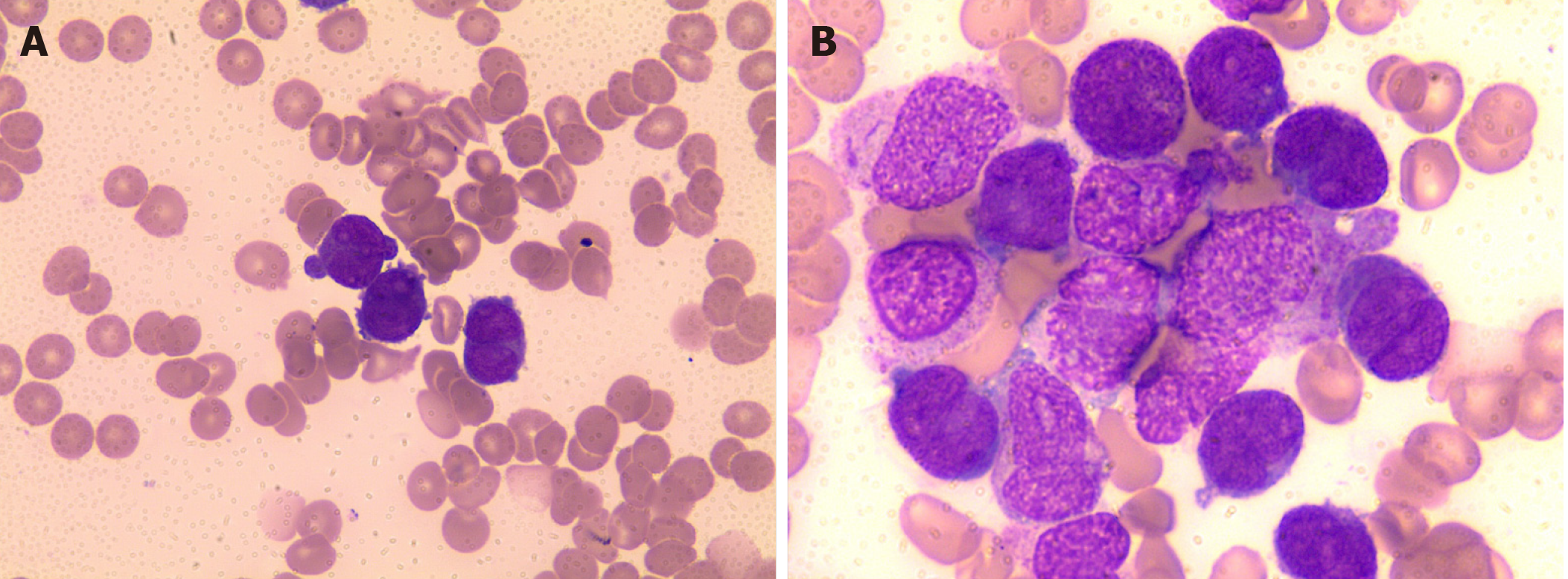
Figure 1 Microscopic examination of peripheral blood and bone marrow.
A: PB showed rouleaux formation of red blood cells and an atypical promyelocyte; B: BM aspiration smears showed increased number of atypical promyelocytes with bilobed nuclei and densely packed large granules in the cytoplasm (Wright-Giemsa staining, × 1000). PB: Peripheral blood; BM: Bone marrow.
- Citation: Hong LL, Sheng XF, Zhuang HF. Therapy-related acute promyelocytic leukemia with FMS-like tyrosine kinase 3-internal tandem duplication mutation in solitary bone plasmacytoma: A case report. World J Clin Cases 2020; 8(19): 4579-4587
- URL: https://www.wjgnet.com/2307-8960/full/v8/i19/4579.htm
- DOI: https://dx.doi.org/10.12998/wjcc.v8.i19.4579


