Copyright
©The Author(s) 2020.
World J Clin Cases. Oct 6, 2020; 8(19): 4565-4571
Published online Oct 6, 2020. doi: 10.12998/wjcc.v8.i19.4565
Published online Oct 6, 2020. doi: 10.12998/wjcc.v8.i19.4565
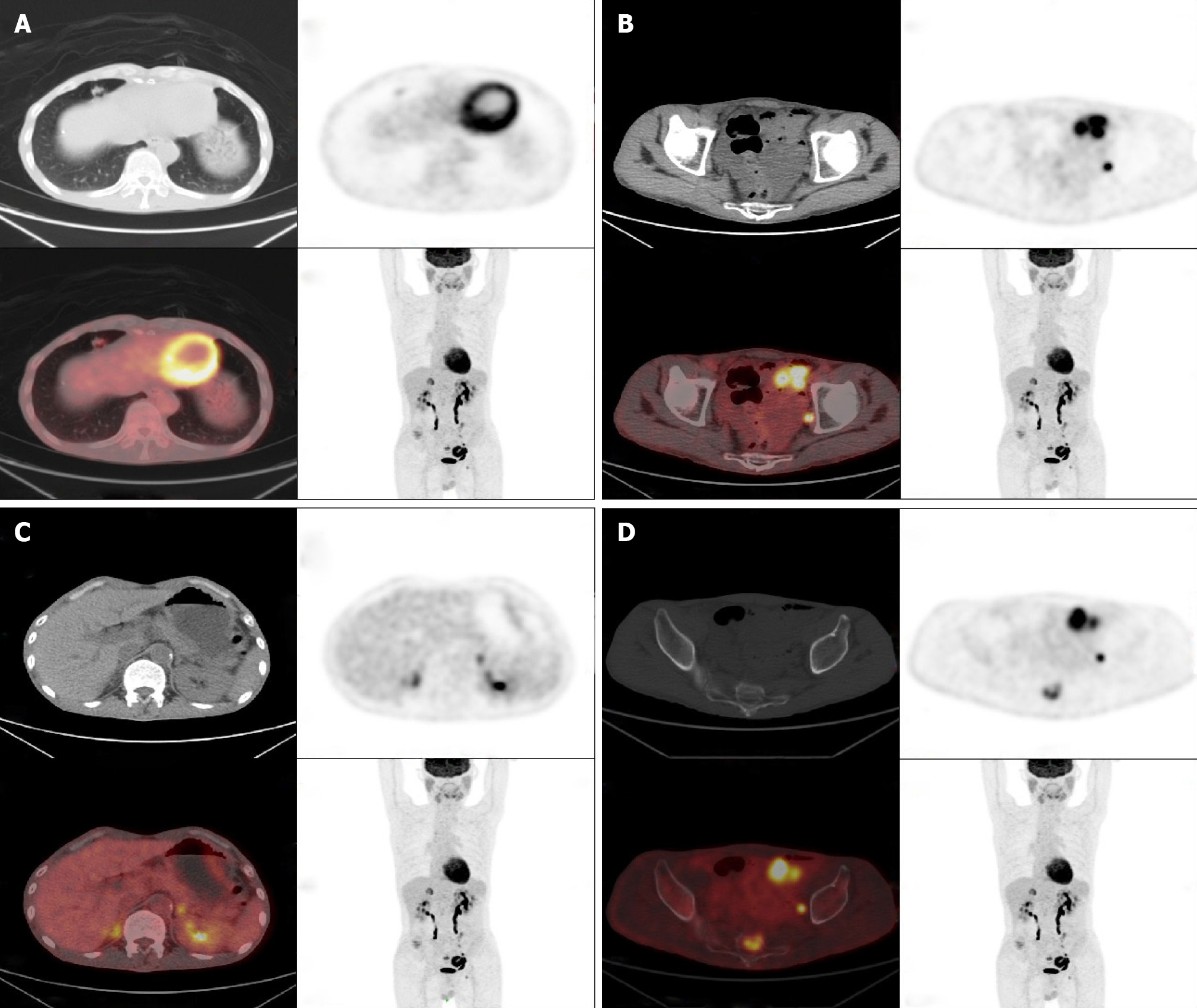
Figure 2 Positron emission computed tomography/ computed tomographic scan.
A: A soft tissue nodule located in the right middle lobe of the lung, about 1.3 cm × 1.0 cm in size, with multiple burrs at the edges and abnormal concentration of tracer; B: Partial small intestinal wall thickness, and abnormal concentration of tracer; C: Bilateral nodular hyperplasia of the adrenal glands, and abnormal concentration of tracer, especially on the right; D: The sacral bone with uneven density, and abnormal concentration of tracer.
- Citation: Hui YY, Zhu LP, Yang B, Zhang ZY, Zhang YJ, Chen X, Wang BM. Gastrointestinal bleeding caused by jejunal angiosarcoma: A case report. World J Clin Cases 2020; 8(19): 4565-4571
- URL: https://www.wjgnet.com/2307-8960/full/v8/i19/4565.htm
- DOI: https://dx.doi.org/10.12998/wjcc.v8.i19.4565


