Copyright
©The Author(s) 2020.
World J Clin Cases. Oct 6, 2020; 8(19): 4558-4564
Published online Oct 6, 2020. doi: 10.12998/wjcc.v8.i19.4558
Published online Oct 6, 2020. doi: 10.12998/wjcc.v8.i19.4558
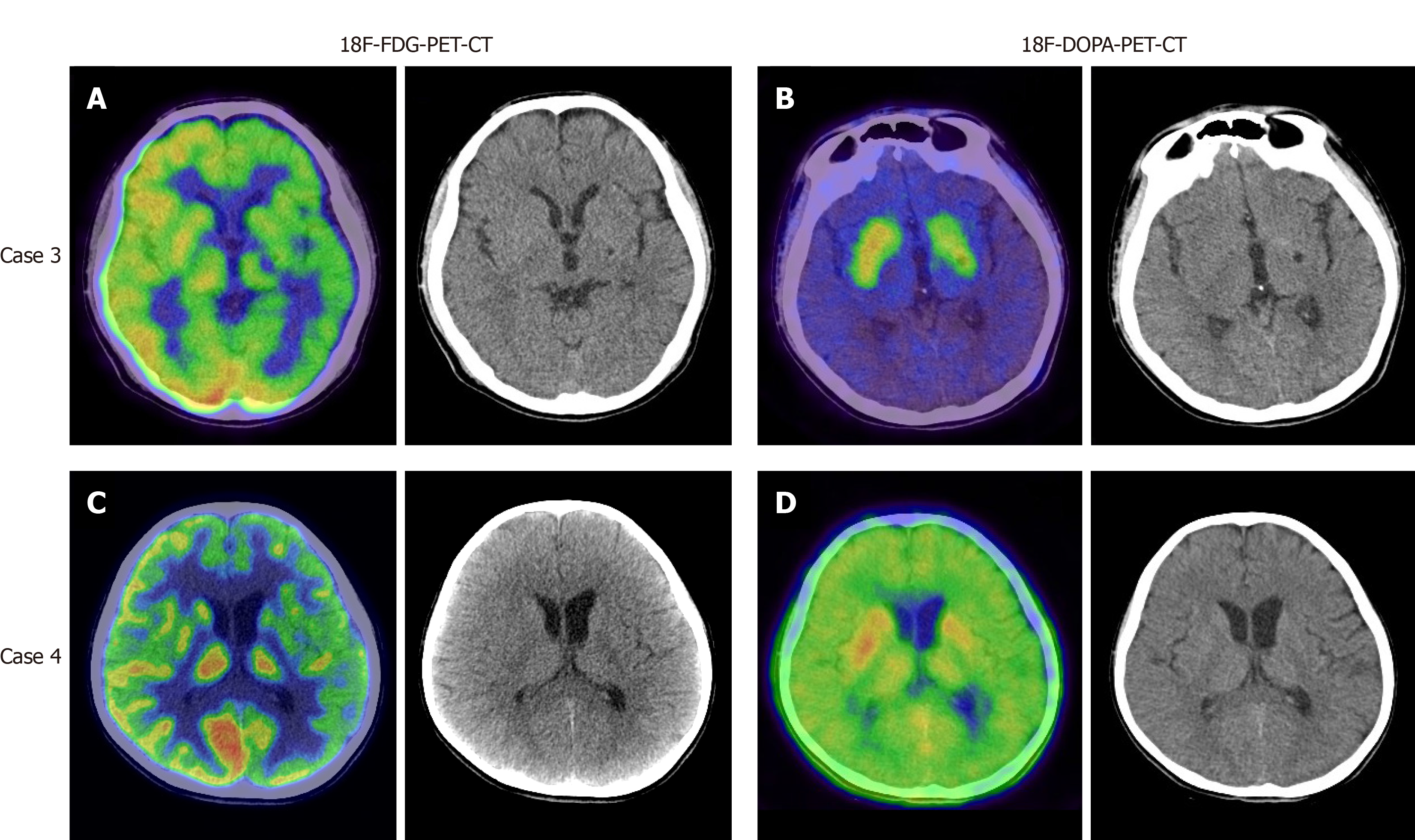
Figure 2 Appearance of germinomas on positron emission tomography-computed tomography.
A: 18F-fluorodeoxyglucose-positron emission tomography-computed tomography (18F-FDG-PET-CT) detected diffuse low FDG uptake in the left hemisphere in case 3; B: 18F-fluorodopa-positron emission tomography-computed tomography (18F-DOPA-PET-CT) detected low uptake of DOPA in the left basal ganglia in case 3; C: 18F-FDG-PET demonstrated low uptake in the left hemisphere in case 4; D: 18F-DOPA-PET-CT showed normal metabolism in case 4. 18F-FDG-PET-CT: 18F-fluorodeoxyglucose-positron emission tomography-computed tomography; 18F-DOPA-PET-CT: 18F-fluorodopa-positron emission tomography-computed tomography.
- Citation: Huang ZC, Dong Q, Song EP, Chen ZJ, Zhang JH, Hou B, Lu ZQ, Qin F. Germinomas of the basal ganglia and thalamus: Four case reports. World J Clin Cases 2020; 8(19): 4558-4564
- URL: https://www.wjgnet.com/2307-8960/full/v8/i19/4558.htm
- DOI: https://dx.doi.org/10.12998/wjcc.v8.i19.4558


