Copyright
©The Author(s) 2020.
World J Clin Cases. Oct 6, 2020; 8(19): 4550-4557
Published online Oct 6, 2020. doi: 10.12998/wjcc.v8.i19.4550
Published online Oct 6, 2020. doi: 10.12998/wjcc.v8.i19.4550
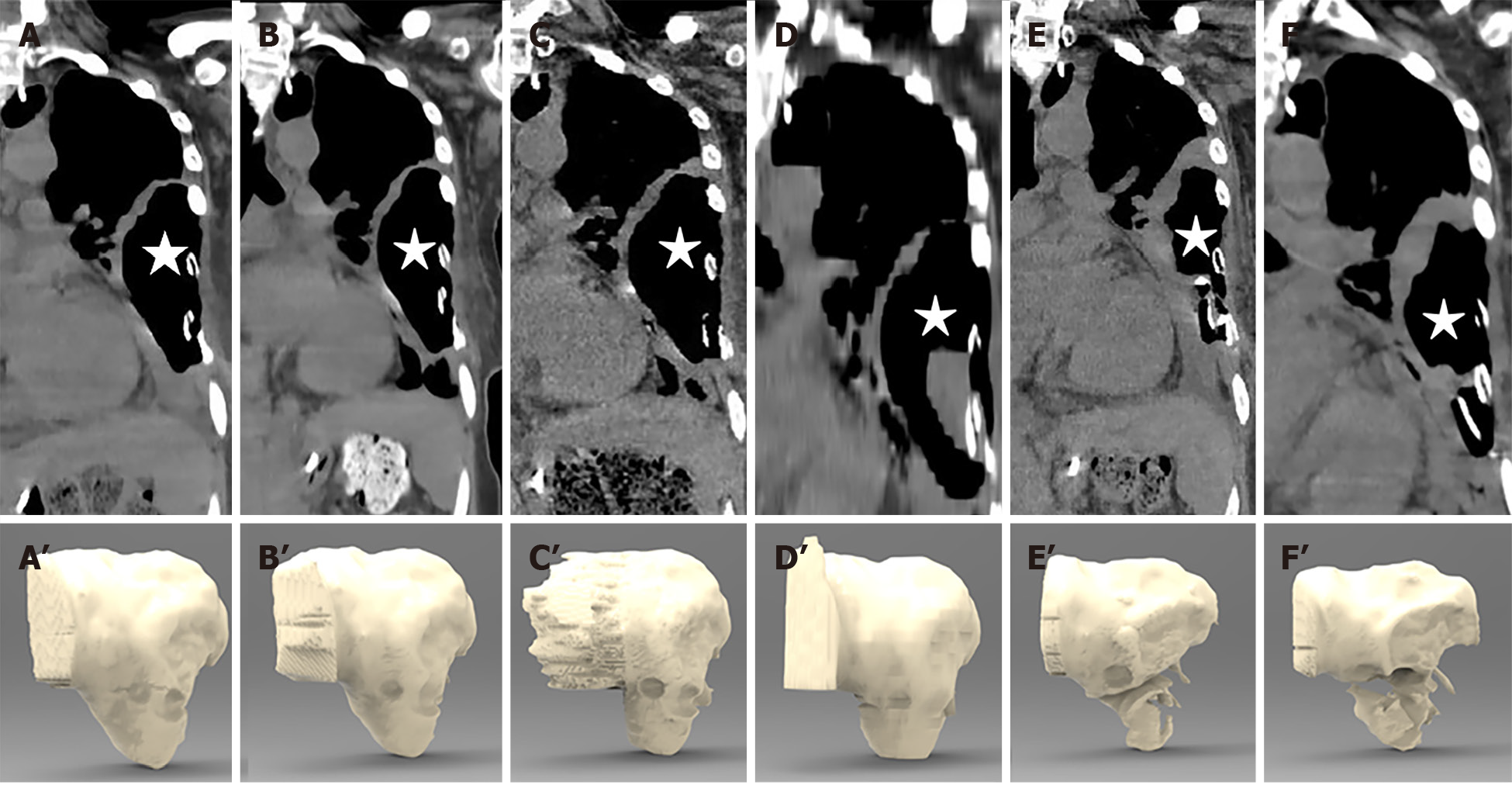
Figure 5 Computed tomography images and 3D stereograms of the gastro-thoracic fistula (asterisk) during the course of treatment.
A-F: Computed tomography examination of the patient on August 20, August 26, August 30, September 2, September 17, and October 8, 2019, respectively, showing that the wall of the thoracic abscess gradually thickened after 2 mo of adequate drainage, together with ozonated water rinse; A’-F’: Three-dimensional stereograms confirming that the volume of the fistula cavity was reduced markedly in late September.
- Citation: Wu DD, Hao KN, Chen XJ, Li XM, He XF. Application of ozonated water for treatment of gastro-thoracic fistula after comprehensive esophageal squamous cell carcinoma therapy: A case report. World J Clin Cases 2020; 8(19): 4550-4557
- URL: https://www.wjgnet.com/2307-8960/full/v8/i19/4550.htm
- DOI: https://dx.doi.org/10.12998/wjcc.v8.i19.4550


