Copyright
©The Author(s) 2020.
World J Clin Cases. Oct 6, 2020; 8(19): 4550-4557
Published online Oct 6, 2020. doi: 10.12998/wjcc.v8.i19.4550
Published online Oct 6, 2020. doi: 10.12998/wjcc.v8.i19.4550
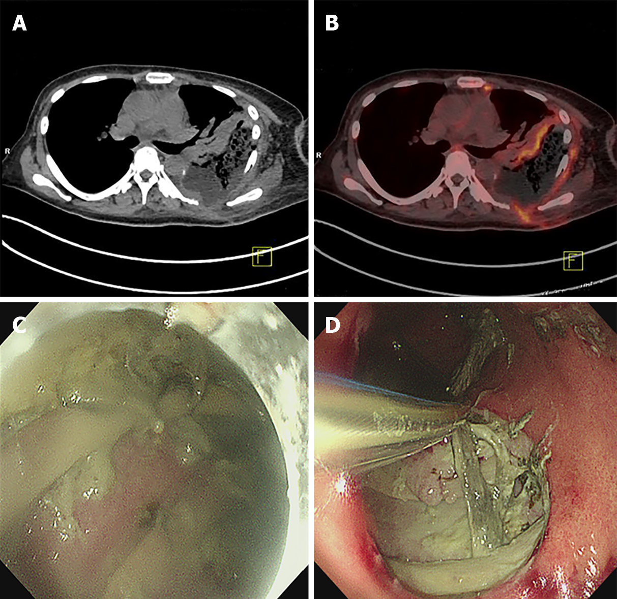
Figure 3 Positron emission tomography-computed tomography and endoscopy examination images of a 50-year-old woman with gastro-thoracic fistula after esophagectomy taken before treatment.
A and B: Computed tomography (CT) and fused CT images showing the formation of a gastro-thoracic fistula, with lesions involving the lateral chest wall; C and D: Endoscopic images showing the purulent gastric contents and local necrosis.
- Citation: Wu DD, Hao KN, Chen XJ, Li XM, He XF. Application of ozonated water for treatment of gastro-thoracic fistula after comprehensive esophageal squamous cell carcinoma therapy: A case report. World J Clin Cases 2020; 8(19): 4550-4557
- URL: https://www.wjgnet.com/2307-8960/full/v8/i19/4550.htm
- DOI: https://dx.doi.org/10.12998/wjcc.v8.i19.4550


