Copyright
©The Author(s) 2020.
World J Clin Cases. Oct 6, 2020; 8(19): 4512-4520
Published online Oct 6, 2020. doi: 10.12998/wjcc.v8.i19.4512
Published online Oct 6, 2020. doi: 10.12998/wjcc.v8.i19.4512
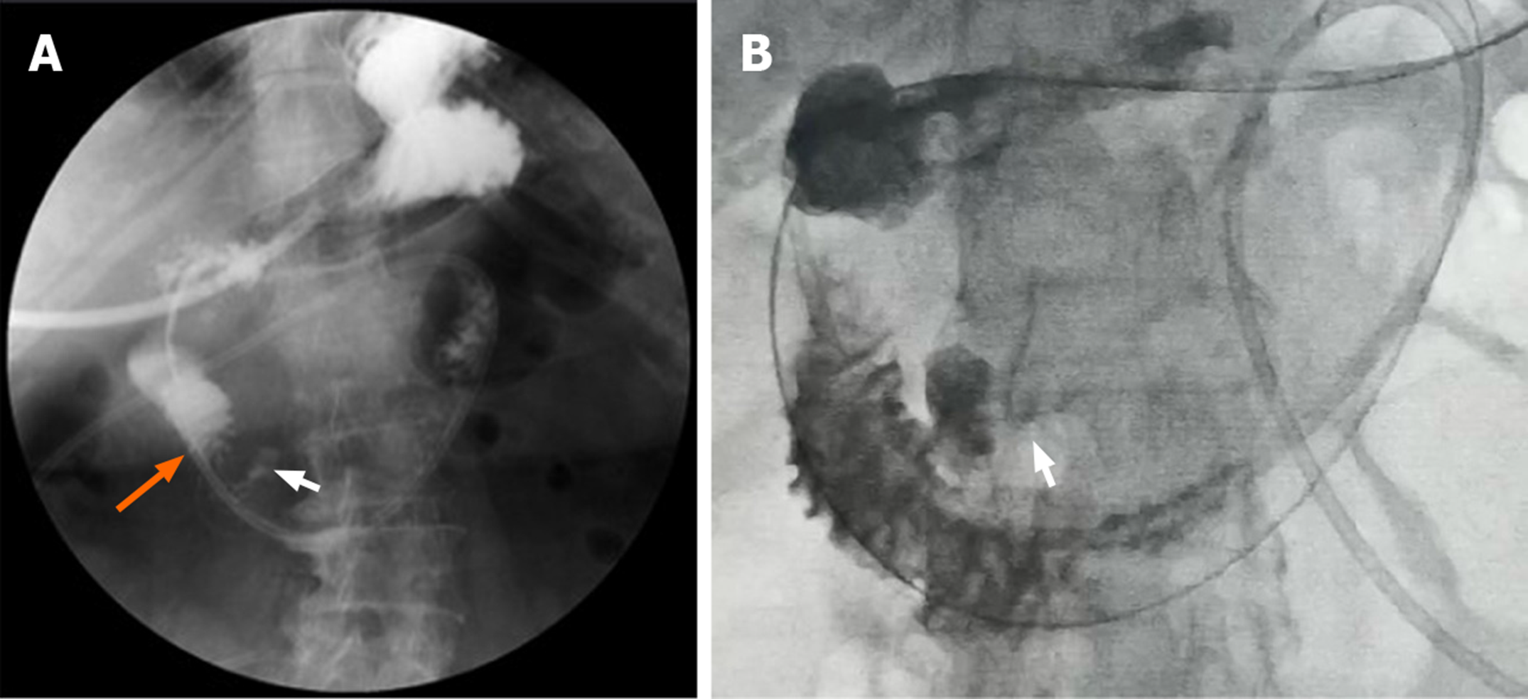
Figure 1 Upper gastrointestinal radiography.
A: Complete stenosis in the descending portion of duodenum (orange arrow) at presentation; B: Resolution of the duodenal obstruction after 3 mo of conservative treatment during the first phase. The white arrow shows the duodenal diverticulum.
- Citation: Zhang BQ, Dai XY, Ye QY, Chang L, Wang ZW, Li XQ, Li YN. Spontaneous resolution of idiopathic intestinal obstruction after pneumonia: A case report. World J Clin Cases 2020; 8(19): 4512-4520
- URL: https://www.wjgnet.com/2307-8960/full/v8/i19/4512.htm
- DOI: https://dx.doi.org/10.12998/wjcc.v8.i19.4512


