Copyright
©The Author(s) 2020.
World J Clin Cases. Oct 6, 2020; 8(19): 4488-4493
Published online Oct 6, 2020. doi: 10.12998/wjcc.v8.i19.4488
Published online Oct 6, 2020. doi: 10.12998/wjcc.v8.i19.4488
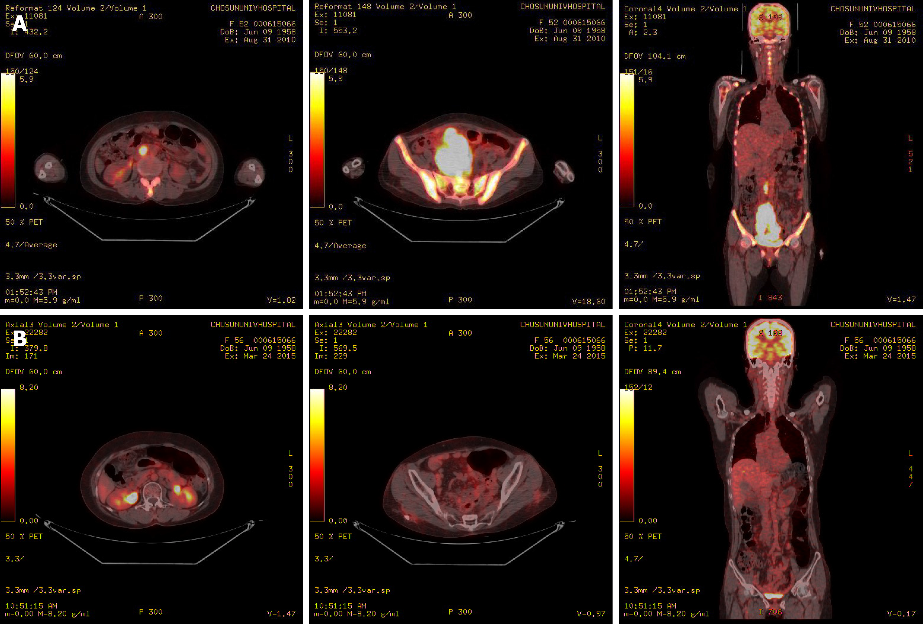
Figure 3 Positron emission tomography–computed tomography.
A: Positron emission tomography–computed tomography revealing hypermetabolism of the huge mass in the pelvic cavity, multiple peritoneal nodules, and para-aortic lymph nodes; B: Hypermetabolism of the huge mass in the pelvic cavity, multiple peritoneal nodules, and para-aortic lymph nodes showing improvement after chemotherapy.
- Citation: Kim HB, Lee HJ, Hong R, Park SG. Extremely rare case of successful treatment of metastatic ovarian undifferentiated carcinoma with high-dose combination cytotoxic chemotherapy: A case report. World J Clin Cases 2020; 8(19): 4488-4493
- URL: https://www.wjgnet.com/2307-8960/full/v8/i19/4488.htm
- DOI: https://dx.doi.org/10.12998/wjcc.v8.i19.4488


