Copyright
©The Author(s) 2020.
World J Clin Cases. Oct 6, 2020; 8(19): 4388-4399
Published online Oct 6, 2020. doi: 10.12998/wjcc.v8.i19.4388
Published online Oct 6, 2020. doi: 10.12998/wjcc.v8.i19.4388
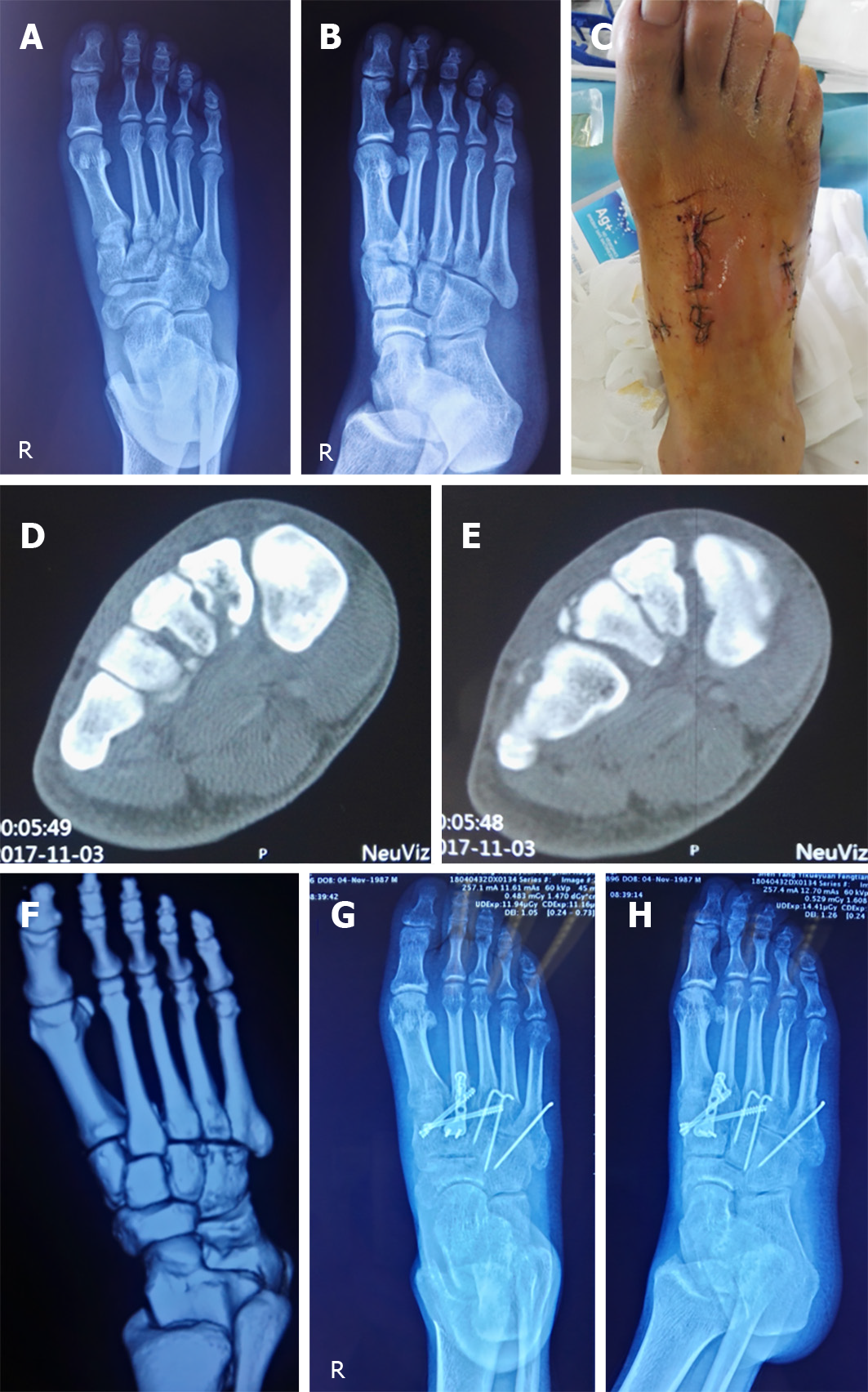
Figure 1 A 30-year-old male with a right foot injury due to a heavy crush.
A and B: X-ray images of the injured foot in the orthophoria and oblique positions, showing the avulsion fracture line of the metatarsal base and the displacement of the fracture; dislocation is not obvious; C: The postoperative incision; D-F: Preoperative computed tomography scans, showing multiple avulsion fracture fragments of the metatarsal base; G and H: X-ray images after operation in the orthophoria and oblique positions.
- Citation: Li X, Jia LS, Li A, Xie X, Cui J, Li GL. Clinical study on the surgical treatment of atypical Lisfranc joint complex injury. World J Clin Cases 2020; 8(19): 4388-4399
- URL: https://www.wjgnet.com/2307-8960/full/v8/i19/4388.htm
- DOI: https://dx.doi.org/10.12998/wjcc.v8.i19.4388


