Copyright
©The Author(s) 2020.
World J Clin Cases. Oct 6, 2020; 8(19): 4349-4359
Published online Oct 6, 2020. doi: 10.12998/wjcc.v8.i19.4349
Published online Oct 6, 2020. doi: 10.12998/wjcc.v8.i19.4349
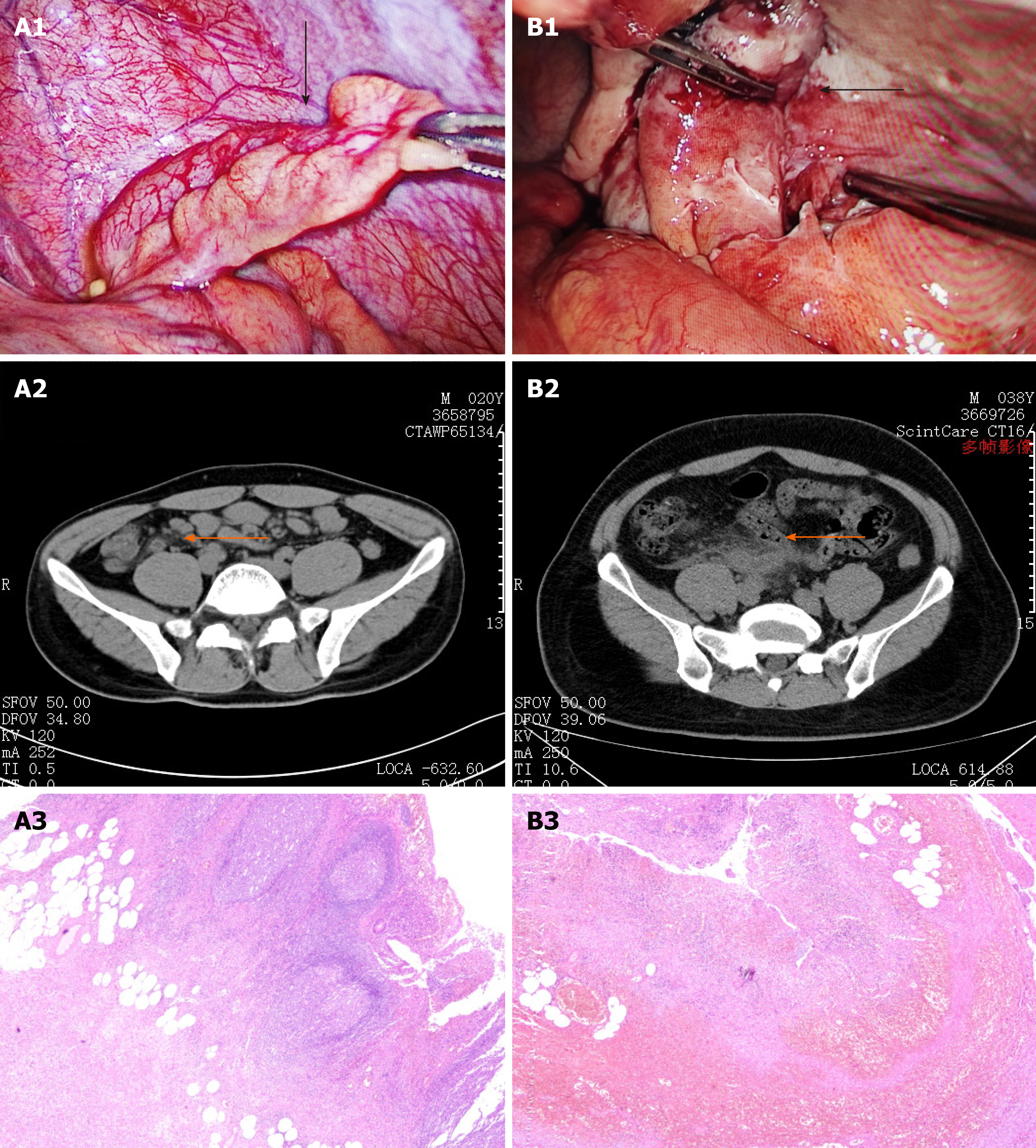
Figure 6 Clinical images from simple group and complex group.
A: Simple group; B: Complex group. Operative photo: A1: Thickened appendix (black arrow) with slightly swollen mesentery; B1: Thickened and congestive appendix (black arrow) with exudation. The distal end of appendix is necrosis with purplish black color. Abdominal computed tomography scan: A2: Slightly thickened appendix (orange arrow) with exudation; B2: Appendix is remarkably thickened and the boundary is obscure. Pathological section (× 40): A3: Structure of appendix remains. The whole layer of appendix indicates obvious infiltration of neutrophils; B3: Structure of appendix disappears. The whole layer of appendix shows hemorrhage and necrosis with infiltration of neutrophils.
- Citation: Zhou Y, Cen LS. Managing acute appendicitis during the COVID-19 pandemic in Jiaxing, China. World J Clin Cases 2020; 8(19): 4349-4359
- URL: https://www.wjgnet.com/2307-8960/full/v8/i19/4349.htm
- DOI: https://dx.doi.org/10.12998/wjcc.v8.i19.4349


