Copyright
©The Author(s) 2020.
World J Clin Cases. Sep 26, 2020; 8(18): 4272-4279
Published online Sep 26, 2020. doi: 10.12998/wjcc.v8.i18.4272
Published online Sep 26, 2020. doi: 10.12998/wjcc.v8.i18.4272
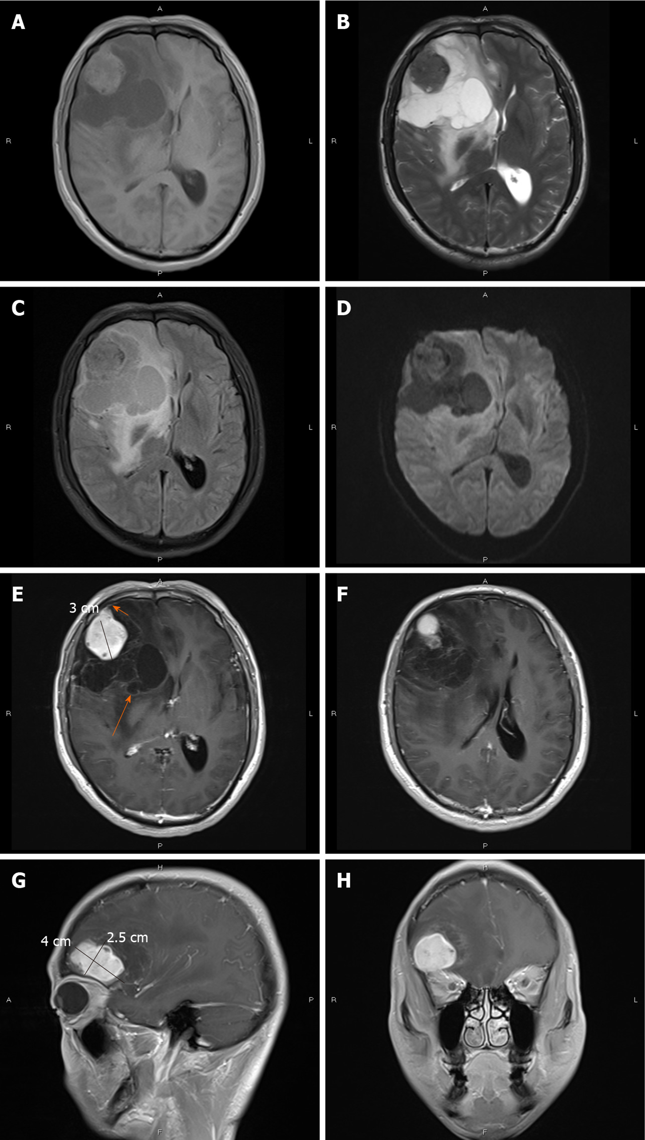
Figure 2 Magnetic resonance imaging findings.
Magnetic resonance imaging showed an irregular, cystic solid mass with surrounding edema in the right frontal lobe. The solid part was approximately 4 cm × 3 cm × 2.5 cm. A: The solid part and cystic wall of the mass exhibited slight hyperintensity on T1-weighted imaging (T1WI); B: Hypointensity on T2-weighted imaging; C: Isointensity on fluid attenuated inversion recovery; D: Diffusion-weighted imaging. Several spots of hypointensity on T1WI and hyperintensity on T2-weighted imaging; E-H: Gadolinium-enhanced T1WI showed that the solid part and cystic wall (long arrow) were strongly enhanced, with dural attachment and dural tail sign (short arrow).
- Citation: Gu KC, Wan Y, Xiang L, Wang LS, Yao WJ. Lymphoplasmacyte-rich meningioma with atypical cystic-solid feature: A case report. World J Clin Cases 2020; 8(18): 4272-4279
- URL: https://www.wjgnet.com/2307-8960/full/v8/i18/4272.htm
- DOI: https://dx.doi.org/10.12998/wjcc.v8.i18.4272


