Copyright
©The Author(s) 2020.
World J Clin Cases. Sep 26, 2020; 8(18): 4272-4279
Published online Sep 26, 2020. doi: 10.12998/wjcc.v8.i18.4272
Published online Sep 26, 2020. doi: 10.12998/wjcc.v8.i18.4272
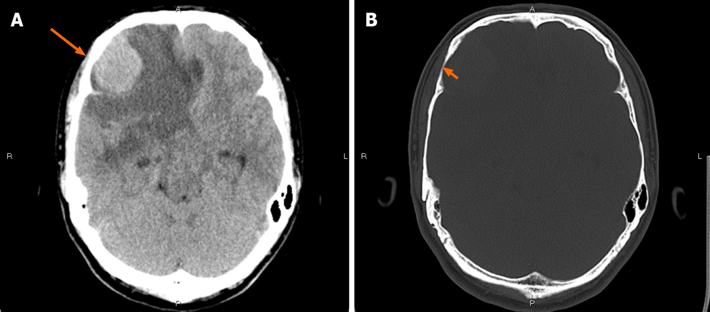
Figure 1 Computed tomography findings.
A: Non-contrast computed tomography showed an irregular hypodense and isodense mixed mass in the right frontal lobe (long arrow), with obvious peritumoral edema around it; B: Bone window showed that the right frontal bone became thin (short arrow).
- Citation: Gu KC, Wan Y, Xiang L, Wang LS, Yao WJ. Lymphoplasmacyte-rich meningioma with atypical cystic-solid feature: A case report. World J Clin Cases 2020; 8(18): 4272-4279
- URL: https://www.wjgnet.com/2307-8960/full/v8/i18/4272.htm
- DOI: https://dx.doi.org/10.12998/wjcc.v8.i18.4272


