Copyright
©The Author(s) 2020.
World J Clin Cases. Sep 26, 2020; 8(18): 4259-4265
Published online Sep 26, 2020. doi: 10.12998/wjcc.v8.i18.4259
Published online Sep 26, 2020. doi: 10.12998/wjcc.v8.i18.4259
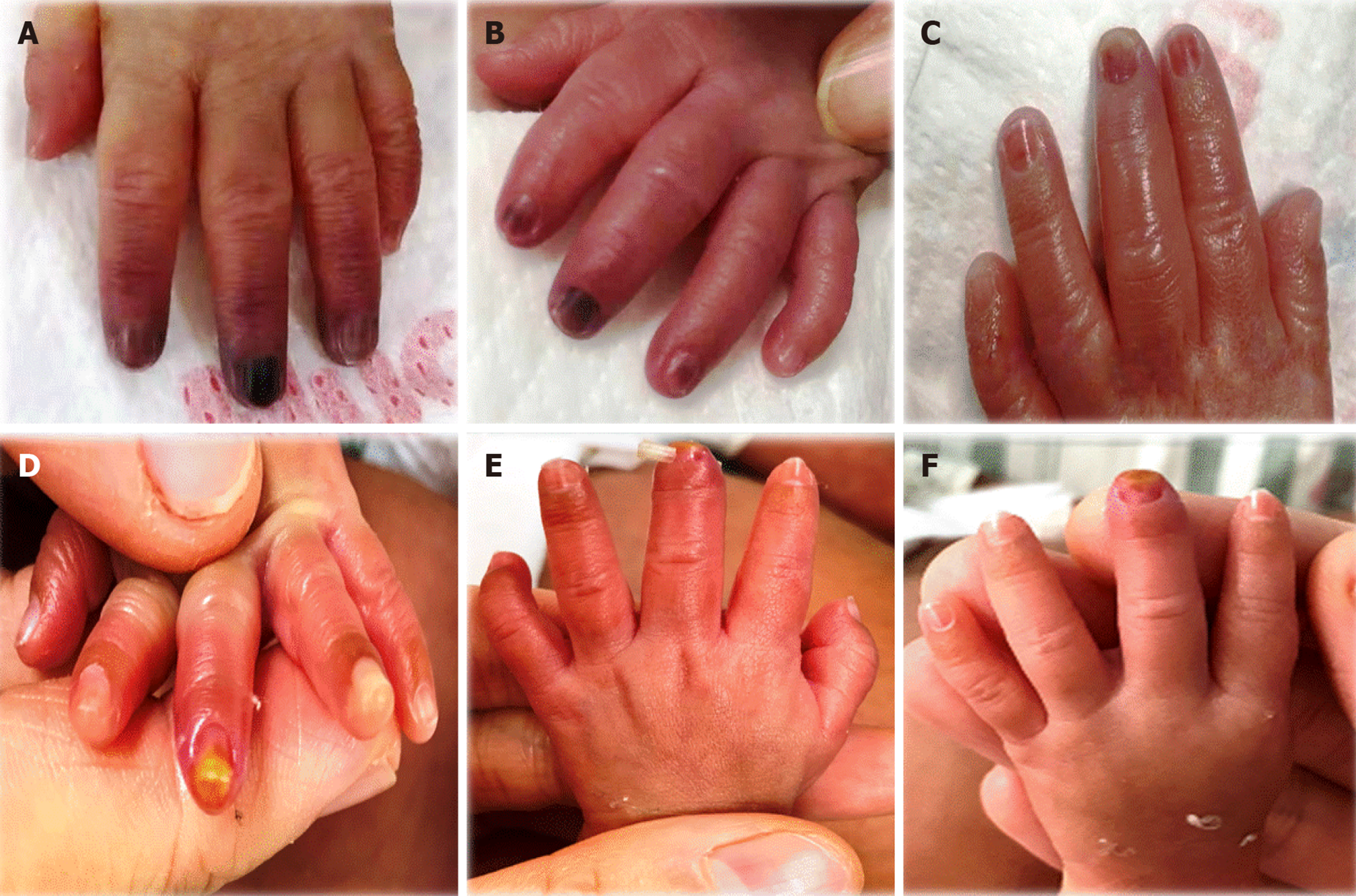
Figure 2 The outcome of left finger-tip artery embolization.
A: On admission, physical examination showed that the palm of the left hand was blue and purple, the left index finger, middle finger, and ring finger end knuckles were black, and the border was unclear. B, C: After a week, the left index finger, middle finger, and ring finger turned ruddy. D: On the 10th day of admission, a yellow-like tissue of approximately 0.5 cm × 0.3 cm was visible on the fingertips. E: On the 13th day of admission, the fingernail of the middle finger was off, with 0.3 cm × 0.5 cm yellow tissue visible at the fingertips. F: On the 19th day of admission, the index finger, middle finger, ring finger of the left hand were rosy, and bruising was visible on the middle fingertips. No exudation or secretions were observed, the degree of recovery reached 95%, and the improvement was obvious.
- Citation: Huang YF, Hu YL, Wan XL, Cheng H, Wu YH, Yang XY, Shi J. Arterial embolism caused by a peripherally inserted central catheter in a very premature infant: A case report and literature review. World J Clin Cases 2020; 8(18): 4259-4265
- URL: https://www.wjgnet.com/2307-8960/full/v8/i18/4259.htm
- DOI: https://dx.doi.org/10.12998/wjcc.v8.i18.4259


