Copyright
©The Author(s) 2020.
World J Clin Cases. Sep 26, 2020; 8(18): 4234-4244
Published online Sep 26, 2020. doi: 10.12998/wjcc.v8.i18.4234
Published online Sep 26, 2020. doi: 10.12998/wjcc.v8.i18.4234
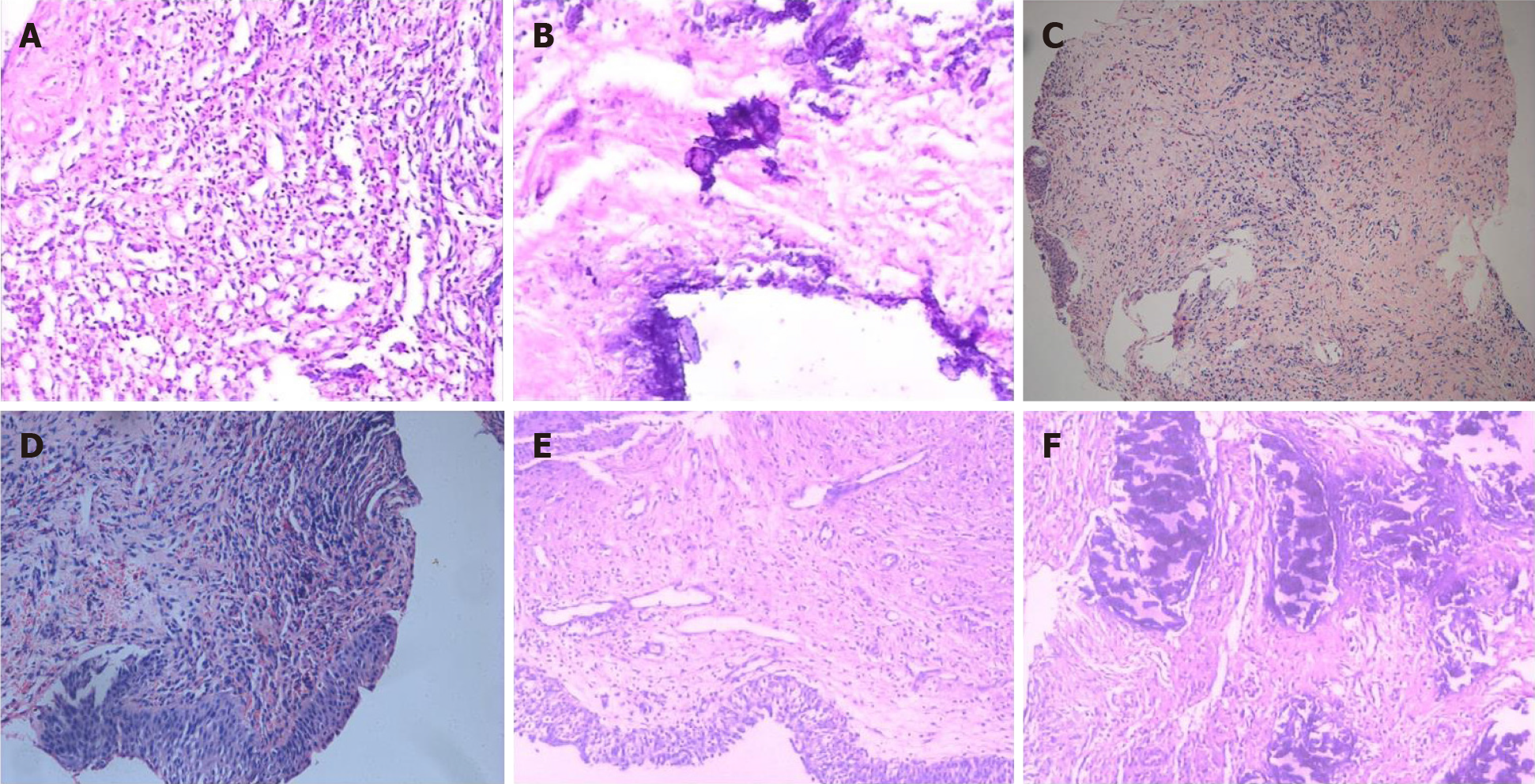
Figure 2 Histopathological examination.
A and B: Hematoxylin and eosin staining on day 22 after onset of encrusted cystitis showed inflammatory granulation tissue on the bladder wall with local necrosis and calcification (100 × and 200 ×, respectively); C and D: Hematoxylin and eosin staining on day 45 after onset of encrusted cystitis showed chronic inflammation and epithelial hyperplasia of bladder mucosa. Calcium deposition, proliferation of fibrous tissue, and infiltration of neutrophils, eosinophils and lymphocytes were observed in the lamina propria of the bladder mucosa (100 × and 200 ×, respectively); E and F: Hematoxylin and eosin staining on day 60 after onset of encrusted cystitis showed that the bladder mucosa was edematous and necrotic with many encrusted crystals and a polymorphonuclear infiltrate forming a thick conglomerate (100 × and 200 ×, respectively).
- Citation: Fu JG, Xie KJ. Successful treatment of encrusted cystitis: A case report and review of literature. World J Clin Cases 2020; 8(18): 4234-4244
- URL: https://www.wjgnet.com/2307-8960/full/v8/i18/4234.htm
- DOI: https://dx.doi.org/10.12998/wjcc.v8.i18.4234


