Copyright
©The Author(s) 2020.
World J Clin Cases. Sep 26, 2020; 8(18): 4234-4244
Published online Sep 26, 2020. doi: 10.12998/wjcc.v8.i18.4234
Published online Sep 26, 2020. doi: 10.12998/wjcc.v8.i18.4234
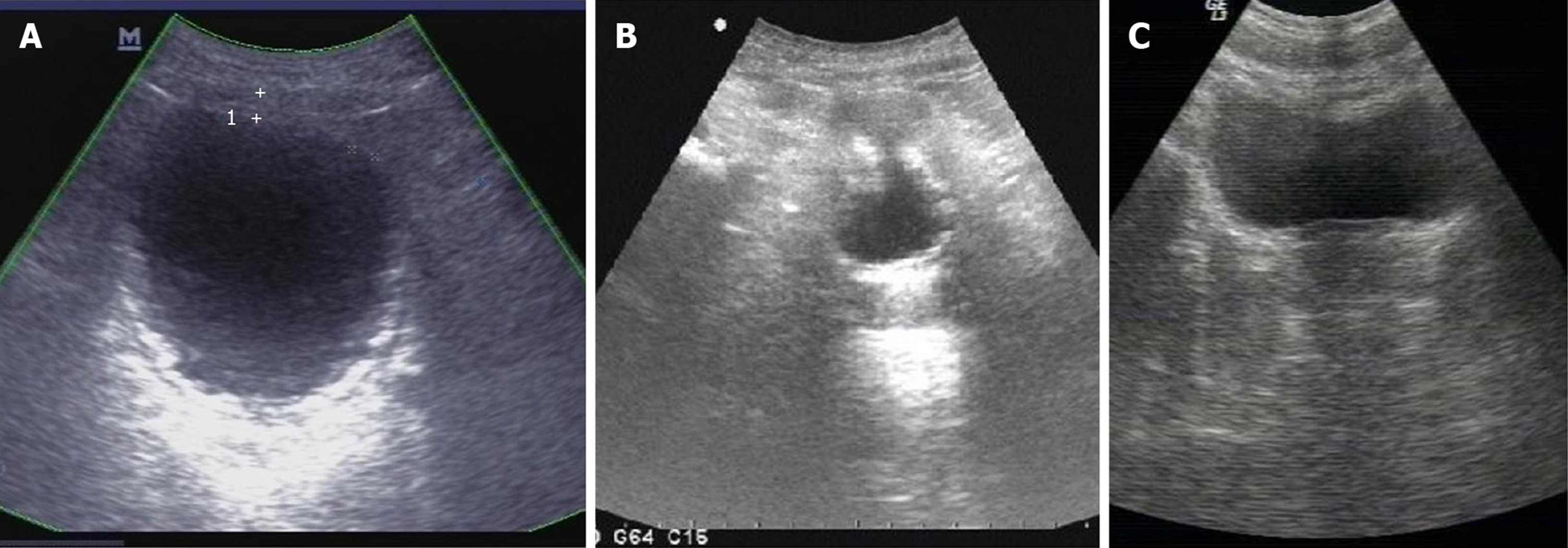
Figure 1 Ultrasonography.
A: Ultrasonography on day 22 after onset of encrusted cystitis showed that the bladder wall was thickened and rough with multiple strong echogenic spots attached to it; B: Ultrasonography on day 60 after onset of encrusted cystitis showed that the bladder wall was thickened (thickest part-9 mm) and rough with multiple strong echogenic spots attached to the wall; C: Ultrasonography on February 8, 2012 showed smooth bladder wall with no hyperechogenic material on it.
- Citation: Fu JG, Xie KJ. Successful treatment of encrusted cystitis: A case report and review of literature. World J Clin Cases 2020; 8(18): 4234-4244
- URL: https://www.wjgnet.com/2307-8960/full/v8/i18/4234.htm
- DOI: https://dx.doi.org/10.12998/wjcc.v8.i18.4234


