Copyright
©The Author(s) 2020.
World J Clin Cases. Sep 26, 2020; 8(18): 4215-4222
Published online Sep 26, 2020. doi: 10.12998/wjcc.v8.i18.4215
Published online Sep 26, 2020. doi: 10.12998/wjcc.v8.i18.4215
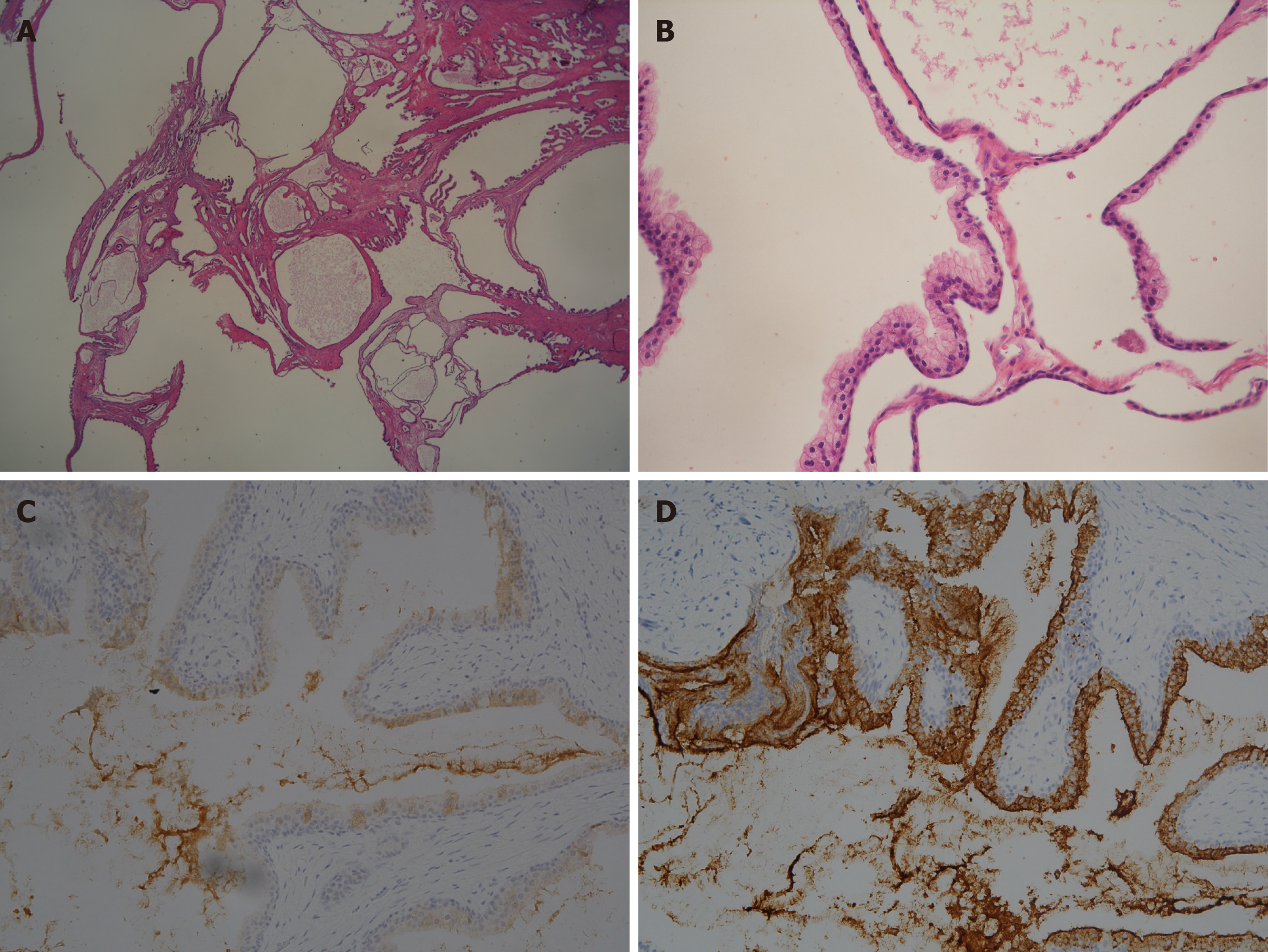
Figure 5 Microscopic findings of the resected tissue.
A: Variously sized dilated glandular and cystic structures lined by blended prostatic type epithelia on low-power magnification (hematoxylin and eosin, original magnification 10 ×); B: Glands and cysts are lined by cuboidal to columnar epithelial cells with a distinct, intact basal cell layer (40 ×); C and D: The epithelium of the cysts stained positively for prostate-specific antigen and weakly expressed prostate-specific membrane antigen (20 ×).
- Citation: Fan LW, Chang YH, Shao IH, Wu KF, Pang ST. Robotic surgery in giant multilocular cystadenoma of the prostate: A rare case report. World J Clin Cases 2020; 8(18): 4215-4222
- URL: https://www.wjgnet.com/2307-8960/full/v8/i18/4215.htm
- DOI: https://dx.doi.org/10.12998/wjcc.v8.i18.4215


