Copyright
©The Author(s) 2020.
World J Clin Cases. Sep 26, 2020; 8(18): 4215-4222
Published online Sep 26, 2020. doi: 10.12998/wjcc.v8.i18.4215
Published online Sep 26, 2020. doi: 10.12998/wjcc.v8.i18.4215
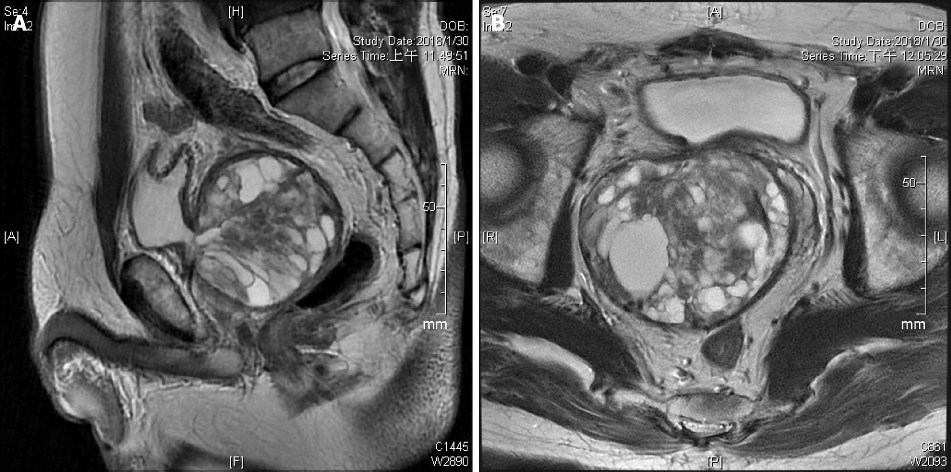
Figure 3 T2-weighted magnetic resonance imaging showing a recurrent cystic multiseptate mass displacing the surrounding structures, (A) sagittal and (B) axial views.
Heterogeneous signal intensity suggests simple fluid, fat, or blood in the cysts. Because of some solid components with low signal intensity in the central part of the tumor, the possibility of a co-existing malignancy could not be eliminated.
- Citation: Fan LW, Chang YH, Shao IH, Wu KF, Pang ST. Robotic surgery in giant multilocular cystadenoma of the prostate: A rare case report. World J Clin Cases 2020; 8(18): 4215-4222
- URL: https://www.wjgnet.com/2307-8960/full/v8/i18/4215.htm
- DOI: https://dx.doi.org/10.12998/wjcc.v8.i18.4215


