Copyright
©The Author(s) 2020.
World J Clin Cases. Sep 26, 2020; 8(18): 4215-4222
Published online Sep 26, 2020. doi: 10.12998/wjcc.v8.i18.4215
Published online Sep 26, 2020. doi: 10.12998/wjcc.v8.i18.4215
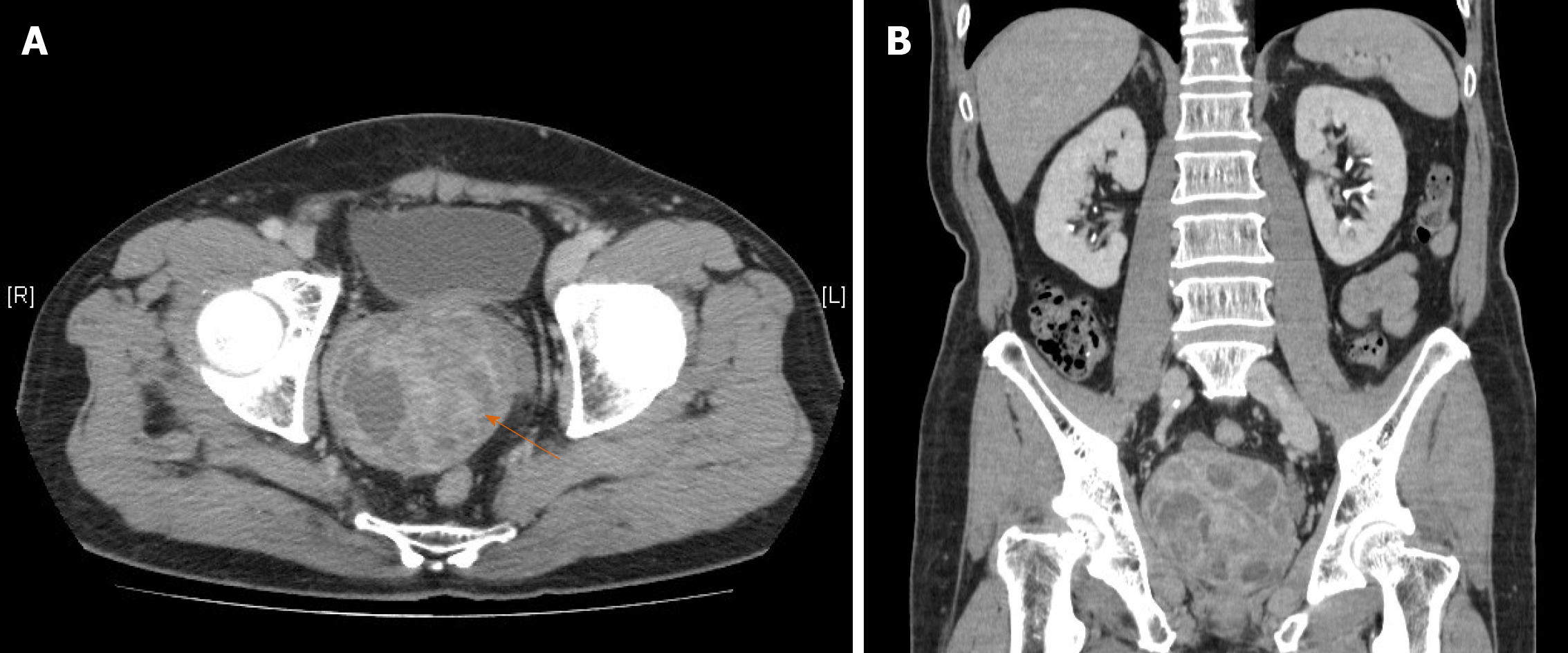
Figure 1 Enhanced computed tomography of the abdomen and pelvis, (A) axial and (B) coronal views, showing a large multiloculated mass with heterogeneous density (arrow) arising from the prostate gland and measuring about 8.
2 cm × 8.1 cm × 7.8 cm in size. Although the tumor occupied almost all the pelvic space, it adhered to the surrounding structures without invasion of the bladder or rectum.
- Citation: Fan LW, Chang YH, Shao IH, Wu KF, Pang ST. Robotic surgery in giant multilocular cystadenoma of the prostate: A rare case report. World J Clin Cases 2020; 8(18): 4215-4222
- URL: https://www.wjgnet.com/2307-8960/full/v8/i18/4215.htm
- DOI: https://dx.doi.org/10.12998/wjcc.v8.i18.4215


