Copyright
©The Author(s) 2020.
World J Clin Cases. Sep 26, 2020; 8(18): 4186-4192
Published online Sep 26, 2020. doi: 10.12998/wjcc.v8.i18.4186
Published online Sep 26, 2020. doi: 10.12998/wjcc.v8.i18.4186
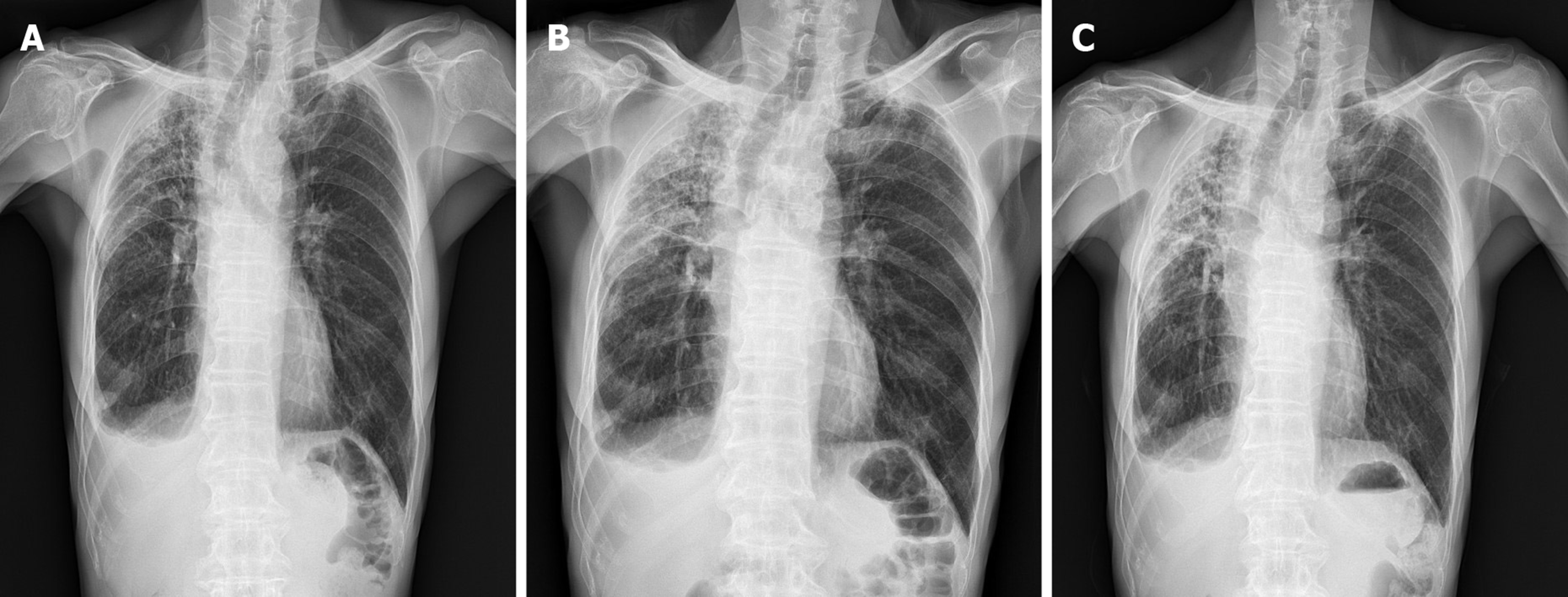
Figure 1 Chest X-ray showed subpleural and irregular shaped increased opacity in right upper lung zone.
Sequential changes during 6 mo showed increased opacity in right upper lung zone. A: Initial visit of diagnosis; B: After 3 mo; C: After 6 mo.
- Citation: Lee JH, Jang HJ, Park JH, Kim HK, Lee S, Kim JY, Kim SH. Unilateral pleuroparenchymal fibroelastosis as a rare form of idiopathic interstitial pneumonia: A case report. World J Clin Cases 2020; 8(18): 4186-4192
- URL: https://www.wjgnet.com/2307-8960/full/v8/i18/4186.htm
- DOI: https://dx.doi.org/10.12998/wjcc.v8.i18.4186


