Copyright
©The Author(s) 2020.
World J Clin Cases. Sep 26, 2020; 8(18): 4128-4134
Published online Sep 26, 2020. doi: 10.12998/wjcc.v8.i18.4128
Published online Sep 26, 2020. doi: 10.12998/wjcc.v8.i18.4128
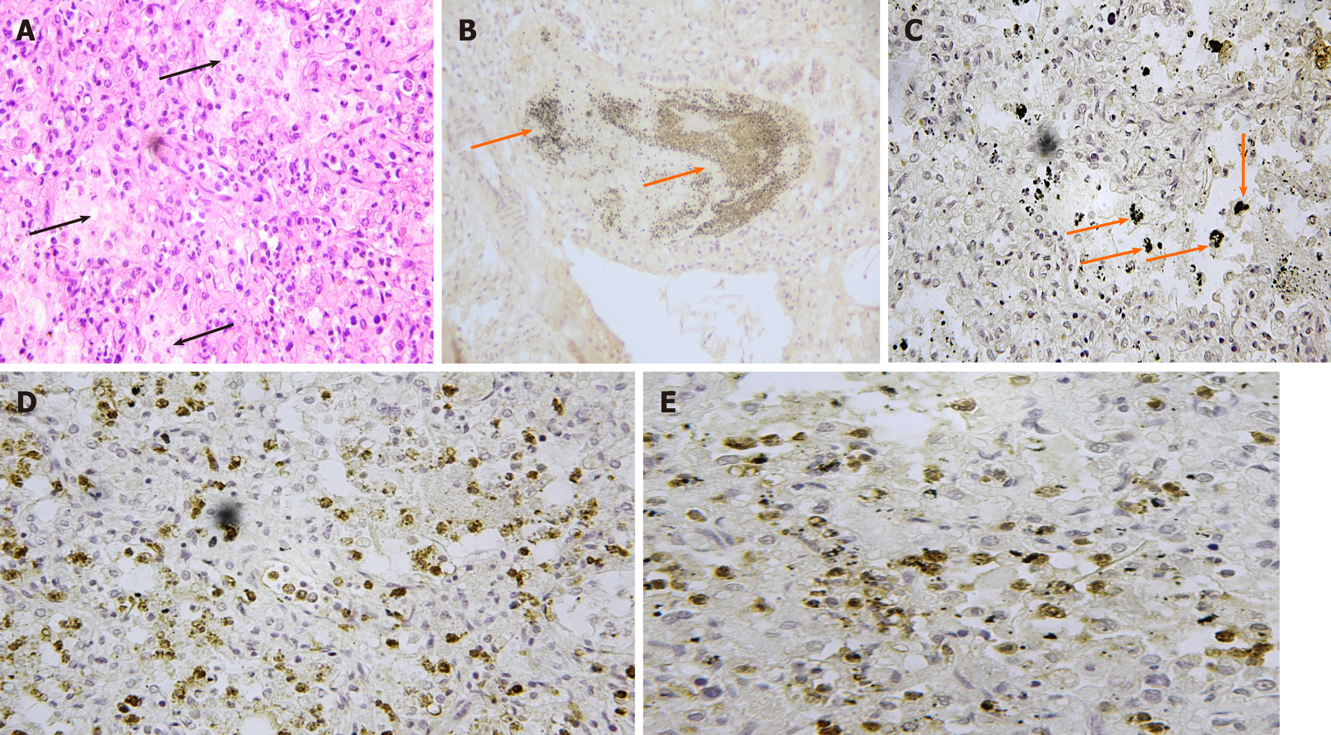
Figure 2 Results of histological examination.
A: Lung tissue, marked congestion of alveolar septal capillaries, which were also engorged with white blood cells, and endo-alveolar incongruous material (black arrows) (haematoxylin-eosin, × 60); B: A positive CD15 mantle of cells (orange arrows) was immersed in endobronchial incongruous material; C: Strong CD68 positivity was visible throughout lung parenchyma (orange arrows); D: Marked alpha-lactalbumin positivity of the intra-alveolar substance was also evident; E: Cytoplasmic staining of macrophages was especially prominent.
- Citation: Maiese A, La Russa R, Arcangeli M, Volonnino G, De Matteis A, Frati P, Fineschi V. Multidisciplinary approach to suspected sudden unexpected infant death caused by milk-aspiration: A case report. World J Clin Cases 2020; 8(18): 4128-4134
- URL: https://www.wjgnet.com/2307-8960/full/v8/i18/4128.htm
- DOI: https://dx.doi.org/10.12998/wjcc.v8.i18.4128


