Copyright
©The Author(s) 2020.
World J Clin Cases. Sep 26, 2020; 8(18): 4094-4099
Published online Sep 26, 2020. doi: 10.12998/wjcc.v8.i18.4094
Published online Sep 26, 2020. doi: 10.12998/wjcc.v8.i18.4094
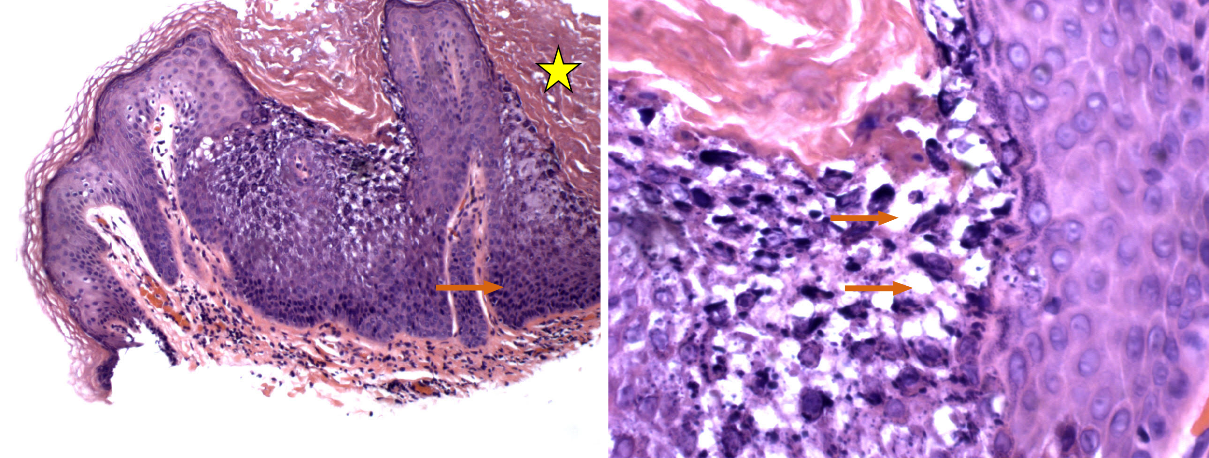
Figure 2 Hematoxylin and eosin photomicrographs of the leison.
The left panel demonstrates compact hyperkeratosis (yellow star) and acanthosis (orange arrow), with the formation of keratin crypts. Higher magnification (right panel) shows intracellular vacuolar degeneration of the epithelial cells of the spinous cell layer, as indicated by the orange arrows.
- Citation: Ginsberg AS, Rajagopalan A, Terlizzi JP. Epidermolytic acanthoma: A case report. World J Clin Cases 2020; 8(18): 4094-4099
- URL: https://www.wjgnet.com/2307-8960/full/v8/i18/4094.htm
- DOI: https://dx.doi.org/10.12998/wjcc.v8.i18.4094


