Copyright
©The Author(s) 2020.
World J Clin Cases. Sep 26, 2020; 8(18): 4094-4099
Published online Sep 26, 2020. doi: 10.12998/wjcc.v8.i18.4094
Published online Sep 26, 2020. doi: 10.12998/wjcc.v8.i18.4094
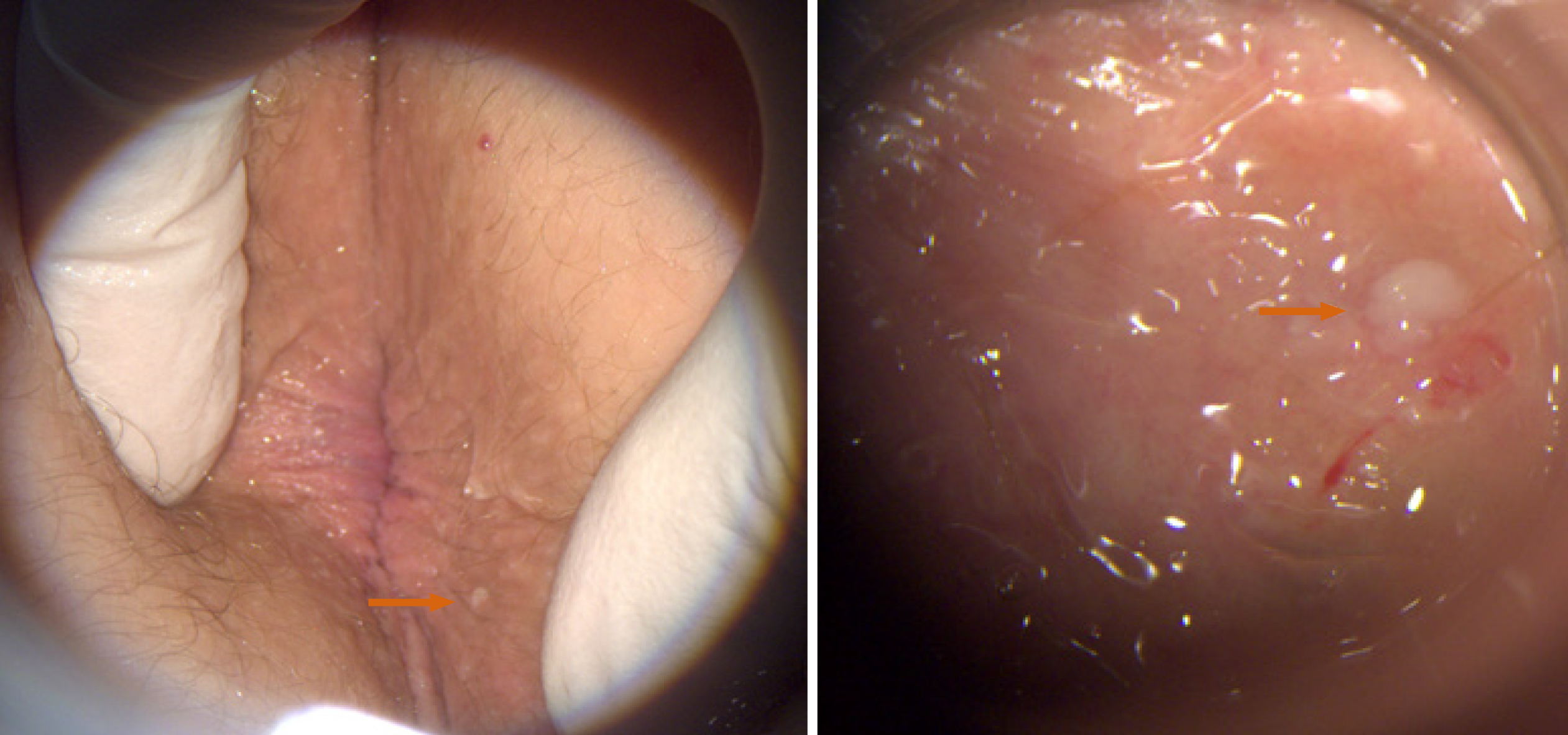
Figure 1 External inspection of the anus demonstrates a pale colored papule three millimeters in size located one centimeter from the anal verge at the right anterior position (left panel).
Magnified view of the lesion with high high resolution anoscopy shows a smooth, well-demarcated perianal lesion (right panel). The right panel shows the same lesion as the left panel at a higher magnification. The magnified photo was taken through a clear anoscope that was used to flatten out the perianal skin folds. The shine is a result of the lubricating gel.
- Citation: Ginsberg AS, Rajagopalan A, Terlizzi JP. Epidermolytic acanthoma: A case report. World J Clin Cases 2020; 8(18): 4094-4099
- URL: https://www.wjgnet.com/2307-8960/full/v8/i18/4094.htm
- DOI: https://dx.doi.org/10.12998/wjcc.v8.i18.4094


