Copyright
©The Author(s) 2020.
World J Clin Cases. Sep 6, 2020; 8(17): 3911-3919
Published online Sep 6, 2020. doi: 10.12998/wjcc.v8.i17.3911
Published online Sep 6, 2020. doi: 10.12998/wjcc.v8.i17.3911
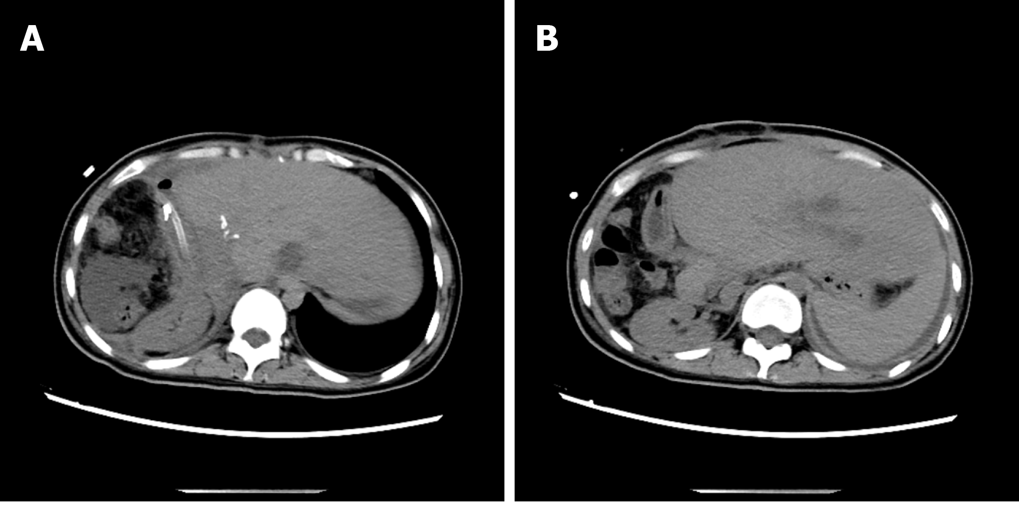
Figure 11 At 3 mo after operation, a small amount of yellowish pus (10-20 mL/24 h) still remained in the abdominal drainage tube (A and B).
Abdominal computed tomography showed that the compensatory increase of the left lobe of liver and the absorption of the peritoneal effusion were obvious. The high-density image in the picture is a metal titanium clip.
- Citation: A JD, Chai JP, Wang H, Gao W, Peng Z, Zhao SY, A XR. Diagnosis and treatment of mixed infection of hepatic cystic and alveolar echinococcosis: Four case reports. World J Clin Cases 2020; 8(17): 3911-3919
- URL: https://www.wjgnet.com/2307-8960/full/v8/i17/3911.htm
- DOI: https://dx.doi.org/10.12998/wjcc.v8.i17.3911


