Copyright
©The Author(s) 2020.
World J Clin Cases. Sep 6, 2020; 8(17): 3911-3919
Published online Sep 6, 2020. doi: 10.12998/wjcc.v8.i17.3911
Published online Sep 6, 2020. doi: 10.12998/wjcc.v8.i17.3911
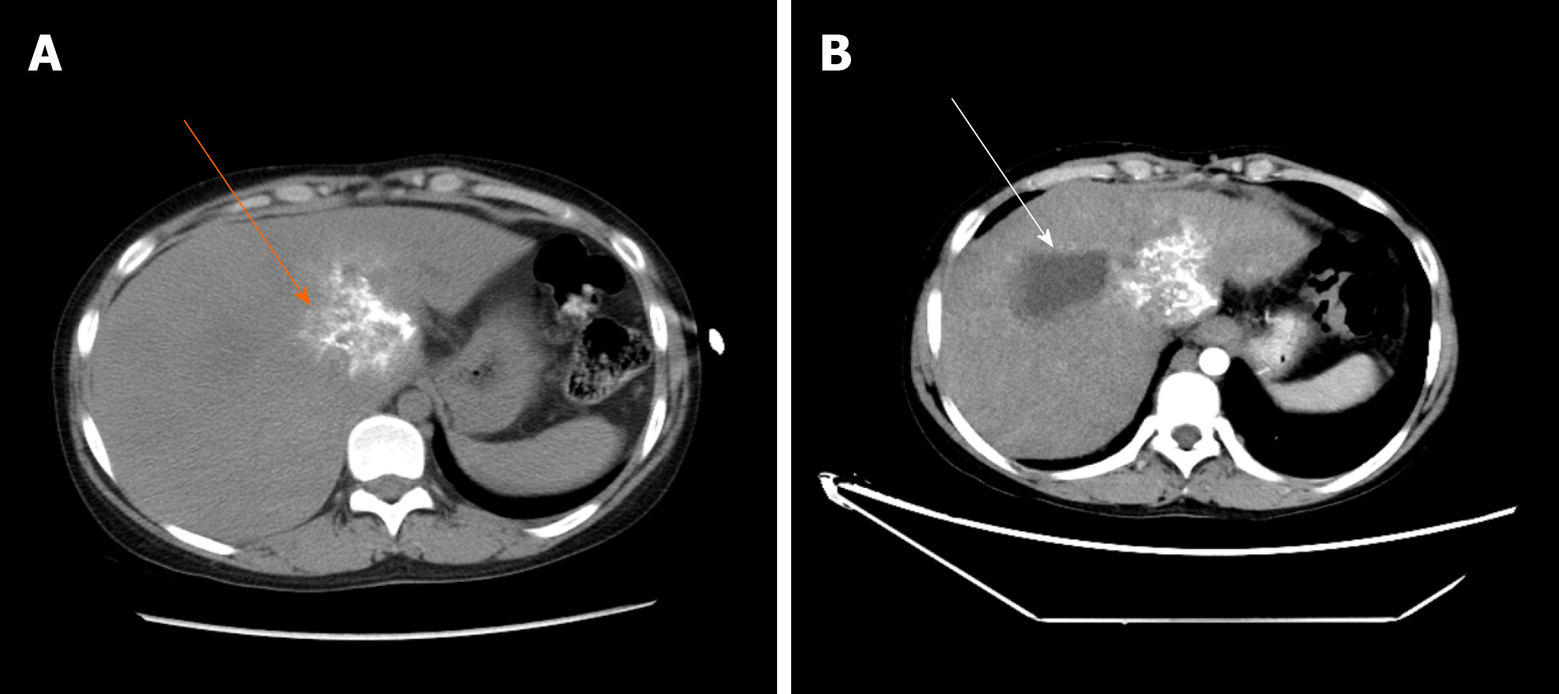
Figure 9 One of the three patients who underwent hepatic hydatid lesion resection again.
A, B: Computed tomography scan 1.5 yr after the operation showed that the lesion of the hepatic S1 segment alveolar echinococcosis still existed, as indicated by the orange arrow. Segments S7 and 8 are the residual cystic cavity of the hydatid cyst, which is indicated by the white arrow; B: The S1 segment of the alveolar hydatid lesions invaded the retrohepatic inferior vena cava. The liver showed congestion-like changes. The blue arrow indicates the retrohepatic inferior vena cava.
- Citation: A JD, Chai JP, Wang H, Gao W, Peng Z, Zhao SY, A XR. Diagnosis and treatment of mixed infection of hepatic cystic and alveolar echinococcosis: Four case reports. World J Clin Cases 2020; 8(17): 3911-3919
- URL: https://www.wjgnet.com/2307-8960/full/v8/i17/3911.htm
- DOI: https://dx.doi.org/10.12998/wjcc.v8.i17.3911


