Copyright
©The Author(s) 2020.
World J Clin Cases. Sep 6, 2020; 8(17): 3881-3889
Published online Sep 6, 2020. doi: 10.12998/wjcc.v8.i17.3881
Published online Sep 6, 2020. doi: 10.12998/wjcc.v8.i17.3881
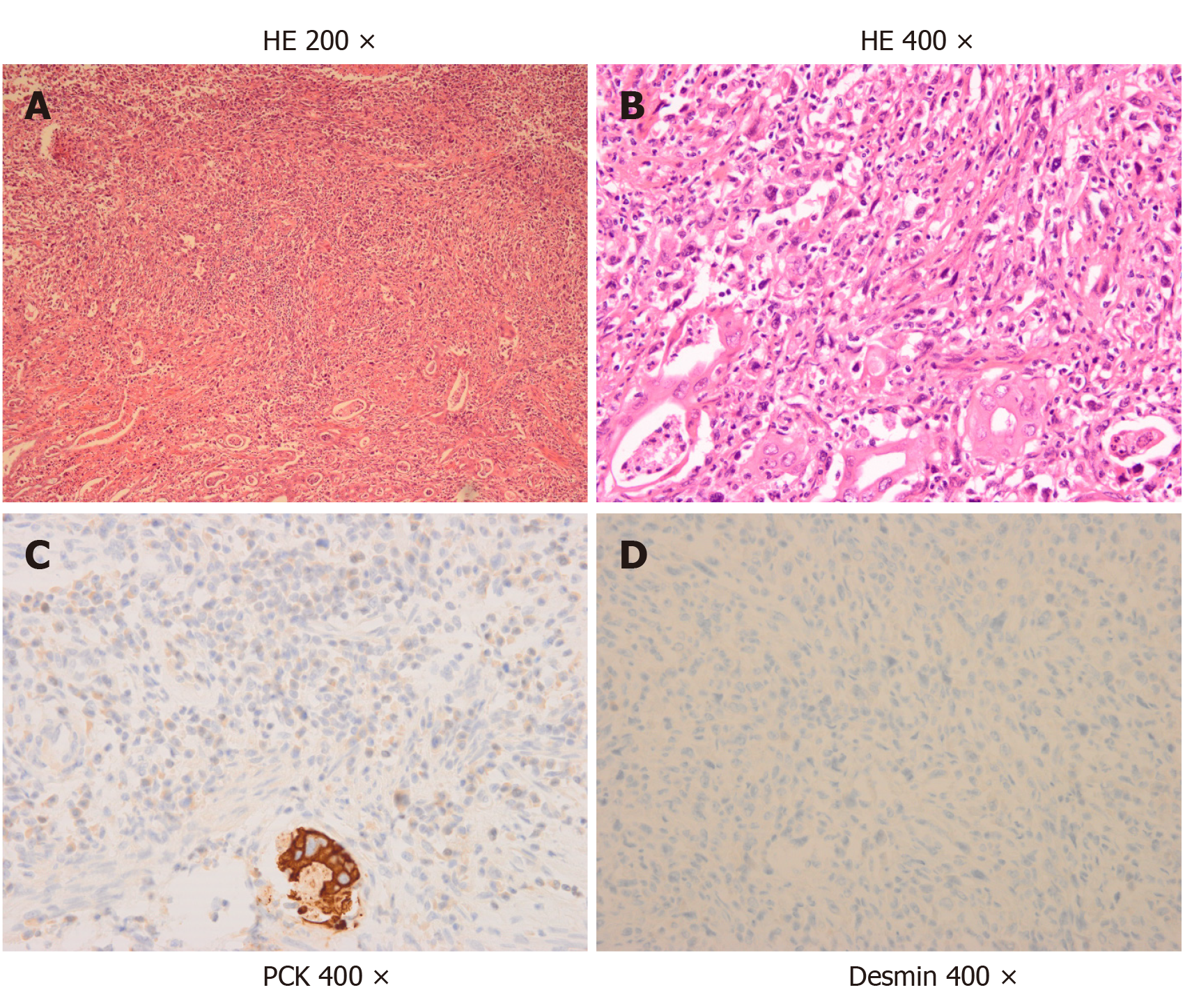
Figure 1 Pathological findings.
A and B: Representative images of hematoxylin and eosin staining (HE)-stained tumor tissue (A: × 200; B: × 400); C and D: Immunohistochemical images showing positive staining for pan-cytokeratin (C) and negative staining for desmin (D) (× 400). HE: Hematoxylin and eosin; PCK: Pan-cytokeratin.
- Citation: Qin Q, Liu M, Wang X. Gallbladder sarcomatoid carcinoma: Seven case reports. World J Clin Cases 2020; 8(17): 3881-3889
- URL: https://www.wjgnet.com/2307-8960/full/v8/i17/3881.htm
- DOI: https://dx.doi.org/10.12998/wjcc.v8.i17.3881


