Copyright
©The Author(s) 2020.
World J Clin Cases. Sep 6, 2020; 8(17): 3853-3858
Published online Sep 6, 2020. doi: 10.12998/wjcc.v8.i17.3853
Published online Sep 6, 2020. doi: 10.12998/wjcc.v8.i17.3853
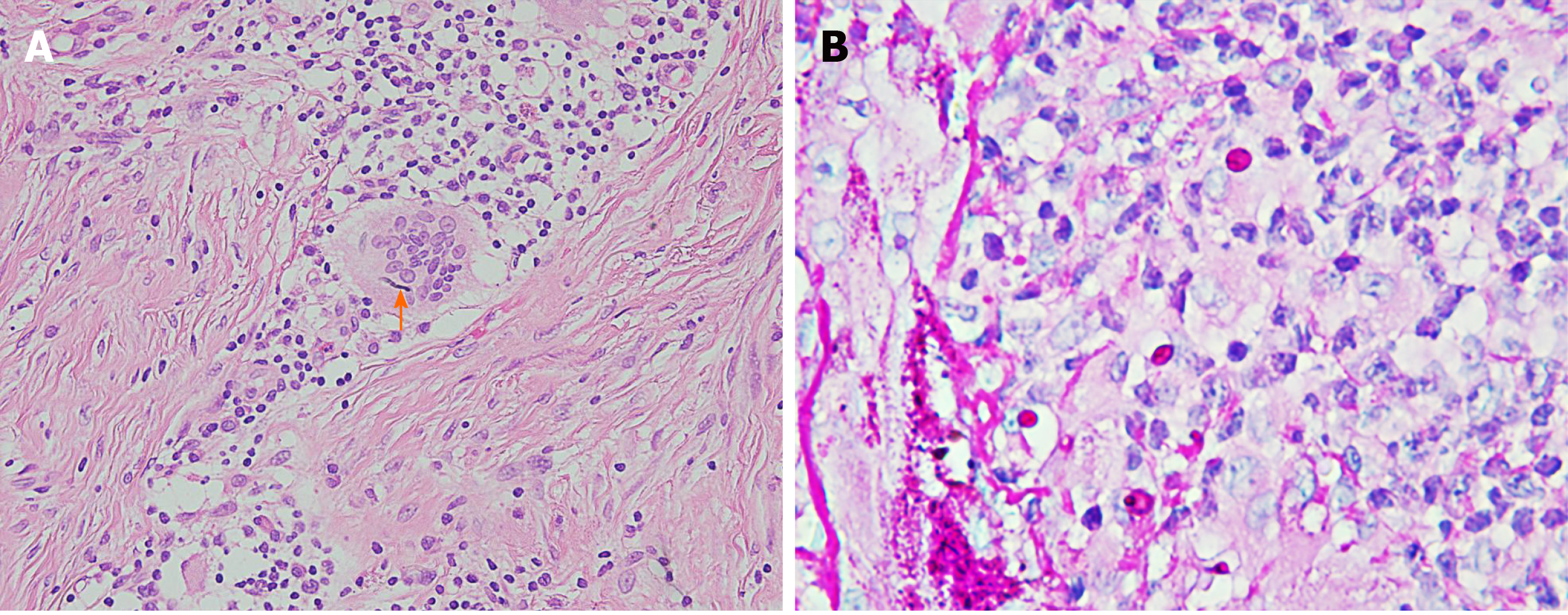
Figure 2 Histologic examination of the skin lesion demonstrated diffuse suppurative and granulomatous inflammation.
The diffuse infiltration of lymphocytes, histiocytes, and multinucleated giant cells was observed in the dermis. A: Fungal mycelium was clearly observed in a giant cell (Hematoxylin-eosin staining, × 400); B: Several giant cells and macrophages were stained with periodic acid-Schiff (Hematoxylin-eosin staining, × 1000).
- Citation: Liu J, Xin WQ, Liu LT, Chen CF, Wu L, Hu XP. Majocchi's granuloma caused by Trichophyton rubrum after facial injection with hyaluronic acid: A case report. World J Clin Cases 2020; 8(17): 3853-3858
- URL: https://www.wjgnet.com/2307-8960/full/v8/i17/3853.htm
- DOI: https://dx.doi.org/10.12998/wjcc.v8.i17.3853


