Copyright
©The Author(s) 2020.
World J Clin Cases. Sep 6, 2020; 8(17): 3835-3840
Published online Sep 6, 2020. doi: 10.12998/wjcc.v8.i17.3835
Published online Sep 6, 2020. doi: 10.12998/wjcc.v8.i17.3835
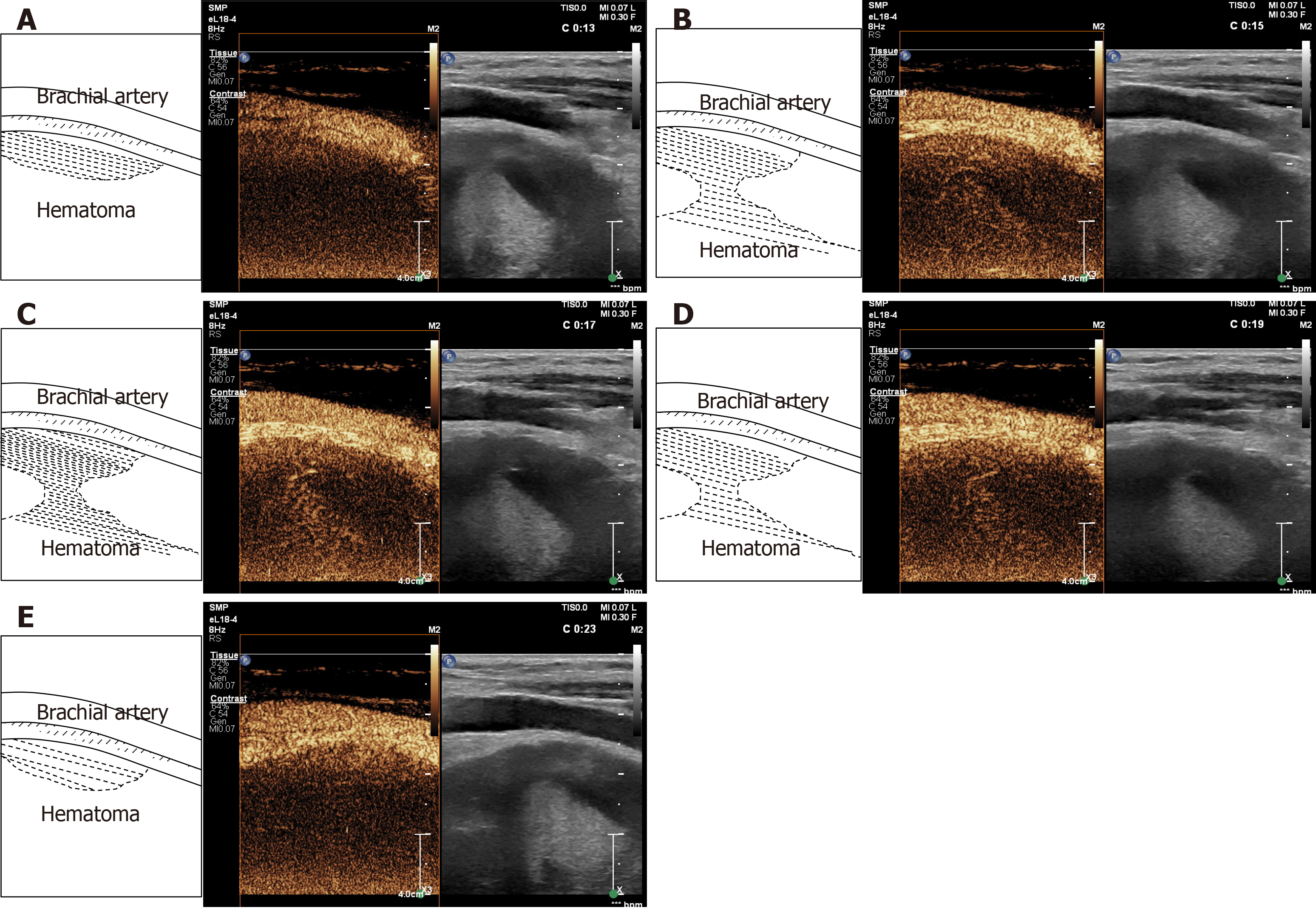
Figure 3 Findings of contrast-enhanced ultrasound.
A: Microbubbles were first detected at 13 s emerging from the brachial artery into the hematoma; B-D: Microbubbles diffused into the hematoma with the local surge of red blood cells; E: Microbubbles disappeared after approximately 23 s.
- Citation: Ma JJ, Zhang B. Diagnosis of an actively bleeding brachial artery hematoma by contrast-enhanced ultrasound: A case report. World J Clin Cases 2020; 8(17): 3835-3840
- URL: https://www.wjgnet.com/2307-8960/full/v8/i17/3835.htm
- DOI: https://dx.doi.org/10.12998/wjcc.v8.i17.3835


