Copyright
©The Author(s) 2020.
World J Clin Cases. Sep 6, 2020; 8(17): 3821-3827
Published online Sep 6, 2020. doi: 10.12998/wjcc.v8.i17.3821
Published online Sep 6, 2020. doi: 10.12998/wjcc.v8.i17.3821
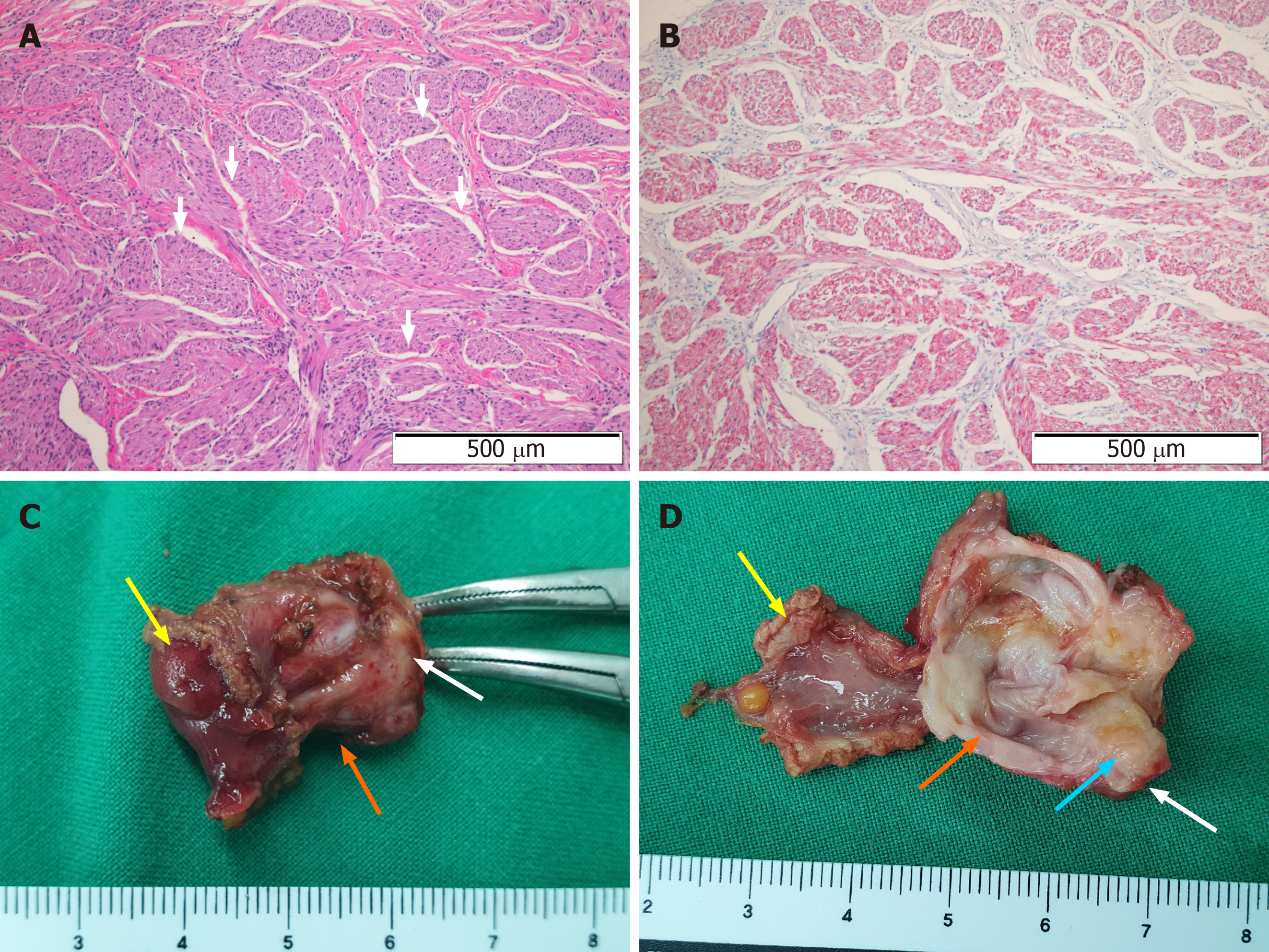
Figure 2 Histological findings of tumor and specimen.
A: Microscopic view of a neuroma showing spindle cell proliferation arranged in short bundles and intervening cleft artifact (white arrows, hematoxylin and eosin staining; magnification x 200); B: Lesion tests positive for S100 protein; C: Macroscopic findings of resected duodenal wall (yellow arrow), lesion (orange arrow), and cystic duct (white arrow); D: Incised specimen showing a hard mass (blue arrow) with the cystic portion (orange arrow) adjacent to the duodenal wall (yellow arrow) and the cystic duct (white arrow).
- Citation: Kim DH, Park JH, Cho JK, Yang JW, Kim TH, Jeong SH, Kim YH, Lee YJ, Hong SC, Jung EJ, Ju YT, Jeong CY, Kim JY. Traumatic neuroma of remnant cystic duct mimicking duodenal subepithelial tumor: A case report. World J Clin Cases 2020; 8(17): 3821-3827
- URL: https://www.wjgnet.com/2307-8960/full/v8/i17/3821.htm
- DOI: https://dx.doi.org/10.12998/wjcc.v8.i17.3821


