Copyright
©The Author(s) 2020.
World J Clin Cases. Sep 6, 2020; 8(17): 3814-3820
Published online Sep 6, 2020. doi: 10.12998/wjcc.v8.i17.3814
Published online Sep 6, 2020. doi: 10.12998/wjcc.v8.i17.3814
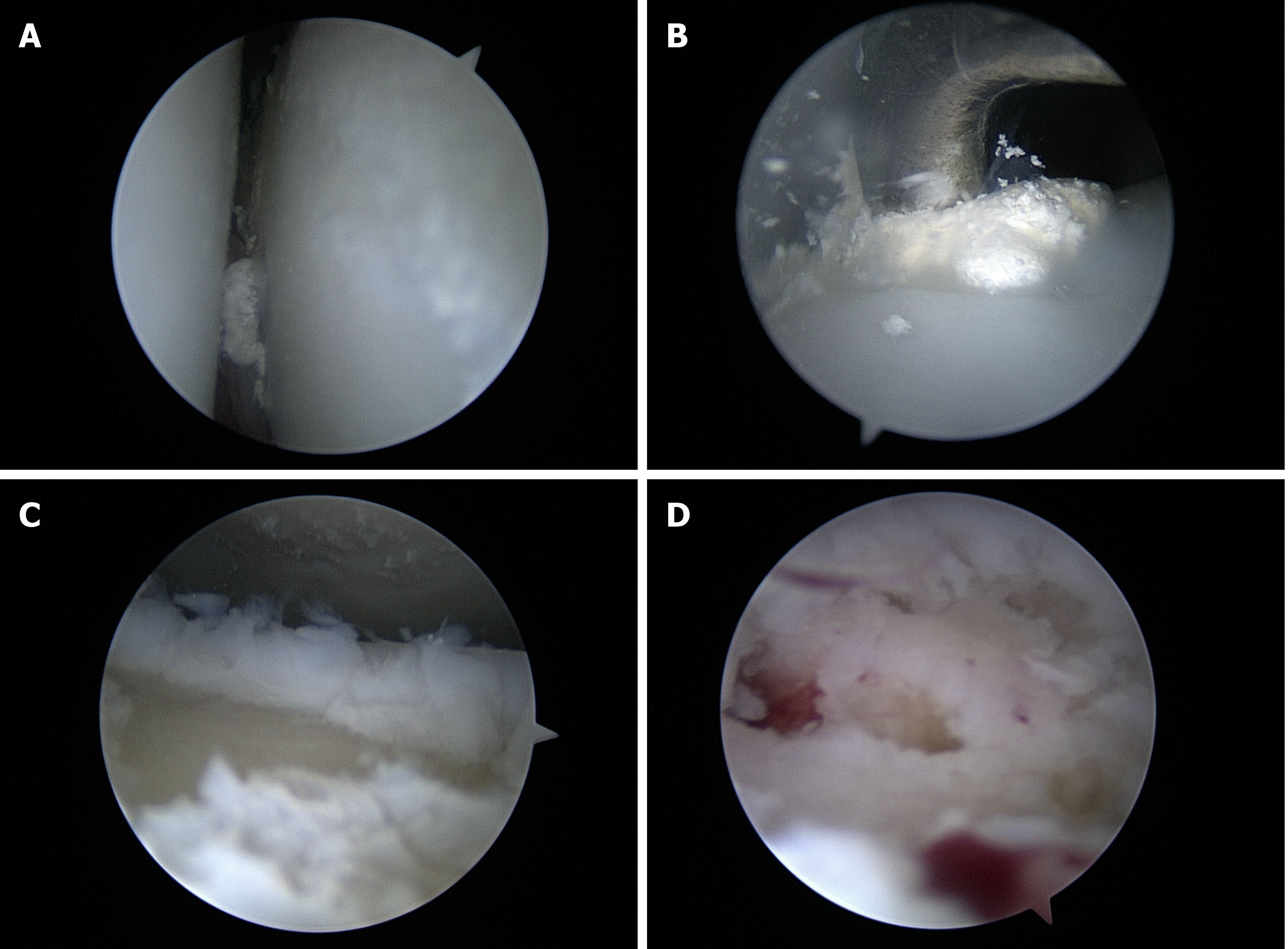
Figure 3 Images of arthroscope were taken during operation.
A: Ankle arthroscopic findings shows tophaceous lesion in the ankle joint; B: Tophaceous lesion was removed. C: Osteochondral defect was checked; D: Microfracture was perfomed.
- Citation: Kim T, Choi YR. Osteochondral lesion of talus with gout tophi deposition: A case report. World J Clin Cases 2020; 8(17): 3814-3820
- URL: https://www.wjgnet.com/2307-8960/full/v8/i17/3814.htm
- DOI: https://dx.doi.org/10.12998/wjcc.v8.i17.3814


