Copyright
©The Author(s) 2020.
World J Clin Cases. Sep 6, 2020; 8(17): 3797-3803
Published online Sep 6, 2020. doi: 10.12998/wjcc.v8.i17.3797
Published online Sep 6, 2020. doi: 10.12998/wjcc.v8.i17.3797
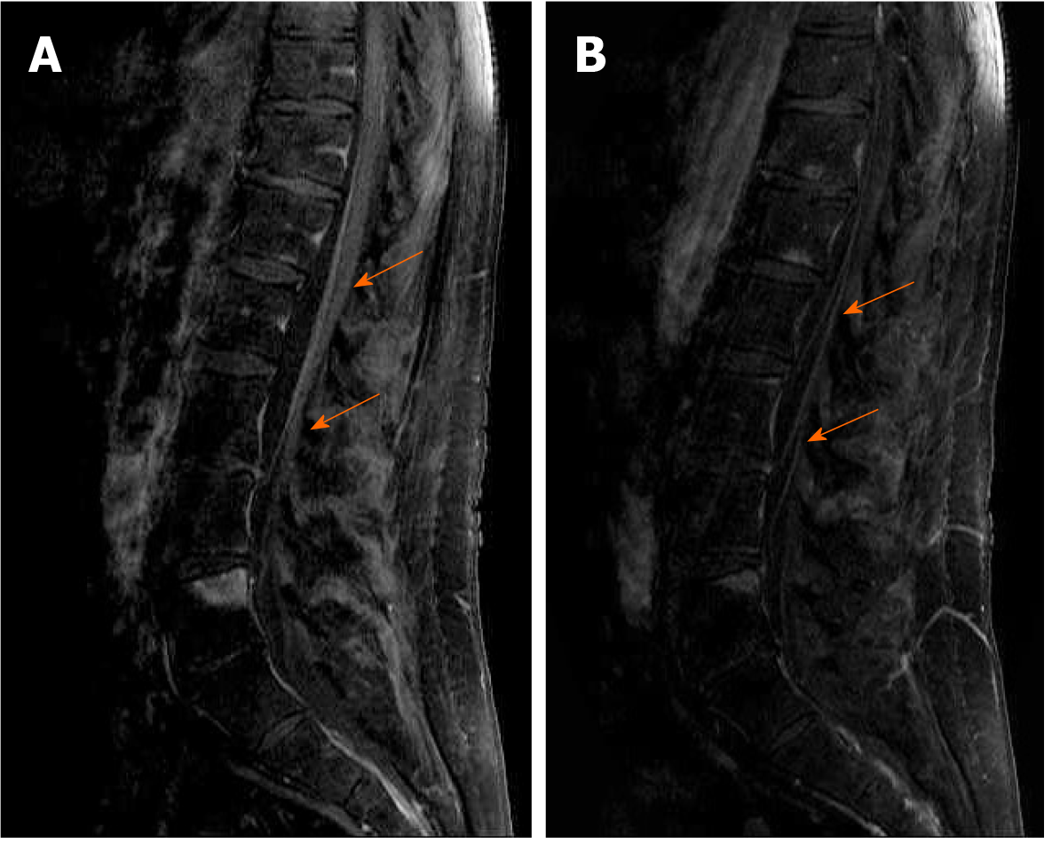
Figure 1 Lumbosacral magnetic resonance imaging scan with intravenous Gadolinium administration in a patient with West Nile virus encephalitis and acute flaccid paresis.
A: Lumbar spine, midsagittal magnetic resonance, contrast-enhanced T1 fat saturation weighted image demonstrated no extrinsic compression, abnormal signal, or enhancement of the lower part of the spinal cord. There was a marked enhancement of the ventral nerve roots of the cauda equina (orange arrows); B: Sagittal contrast-enhanced T1 fat saturation weighted through the region of the cauda equina shows abnormal – prominent enhancement of the nerve roots (orange arrows).
- Citation: Santini M, Zupetic I, Viskovic K, Krznaric J, Kutlesa M, Krajinovic V, Polak VL, Savic V, Tabain I, Barbic L, Bogdanic M, Stevanovic V, Mrzljak A, Vilibic-Cavlek T. Cauda equina arachnoiditis – a rare manifestation of West Nile virus neuroinvasive disease: A case report. World J Clin Cases 2020; 8(17): 3797-3803
- URL: https://www.wjgnet.com/2307-8960/full/v8/i17/3797.htm
- DOI: https://dx.doi.org/10.12998/wjcc.v8.i17.3797


