Copyright
©The Author(s) 2020.
World J Clin Cases. Aug 26, 2020; 8(16): 3591-3600
Published online Aug 26, 2020. doi: 10.12998/wjcc.v8.i16.3591
Published online Aug 26, 2020. doi: 10.12998/wjcc.v8.i16.3591
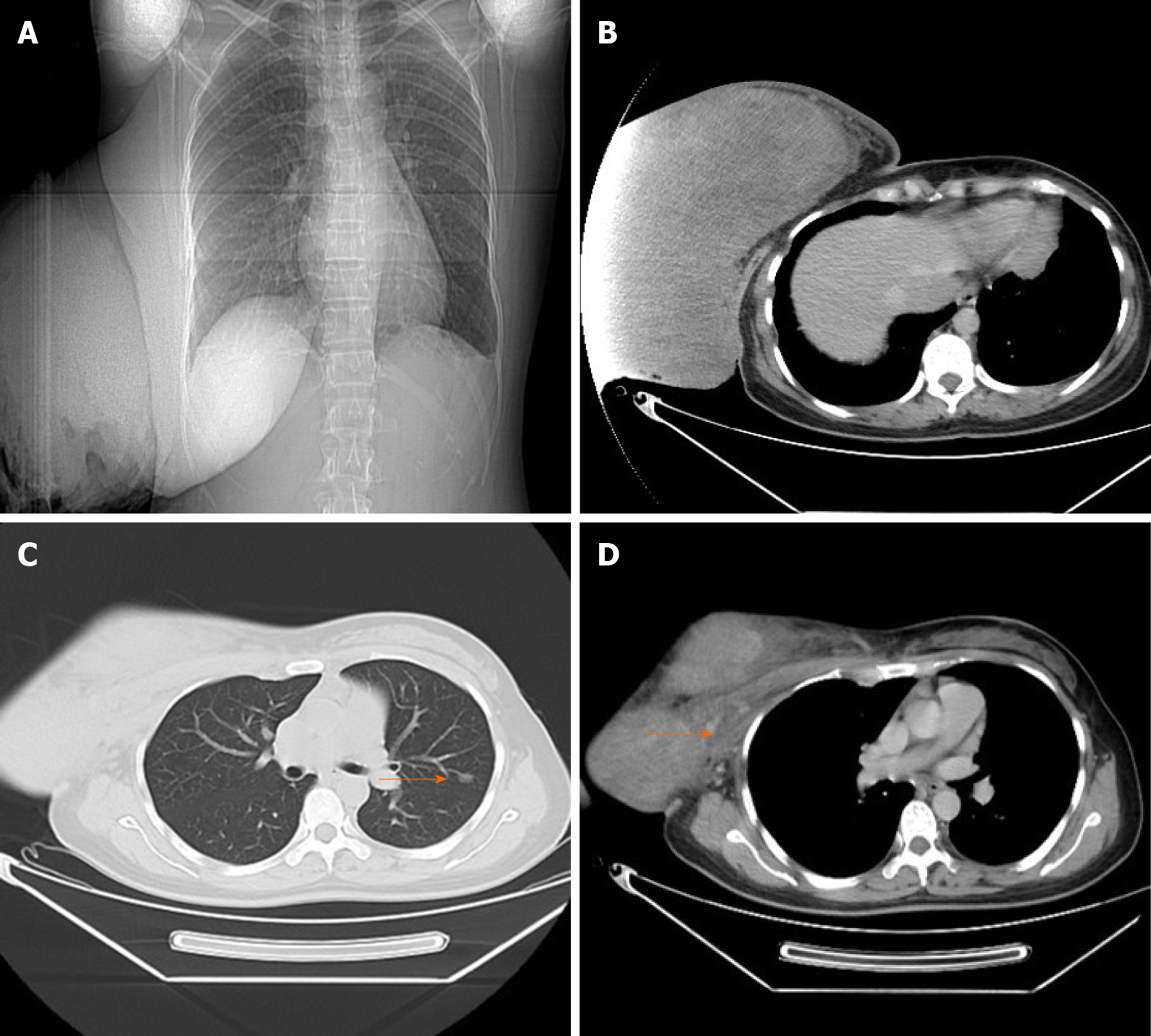
Figure 2 Preoperative radiologic evaluation.
A and B: Chest computed tomography showed a huge mass; C: A pulmonary nodule was seen in the left lung (arrowheads); and D: Preoperative computed tomography showed that there was no clearance between the mass and pectoralis major (arrowheads).
- Citation: Zhang T, Feng L, Lian J, Ren WL. Giant benign phyllodes breast tumour with pulmonary nodule mimicking malignancy: A case report. World J Clin Cases 2020; 8(16): 3591-3600
- URL: https://www.wjgnet.com/2307-8960/full/v8/i16/3591.htm
- DOI: https://dx.doi.org/10.12998/wjcc.v8.i16.3591


