Copyright
©The Author(s) 2020.
World J Clin Cases. Aug 26, 2020; 8(16): 3583-3590
Published online Aug 26, 2020. doi: 10.12998/wjcc.v8.i16.3583
Published online Aug 26, 2020. doi: 10.12998/wjcc.v8.i16.3583
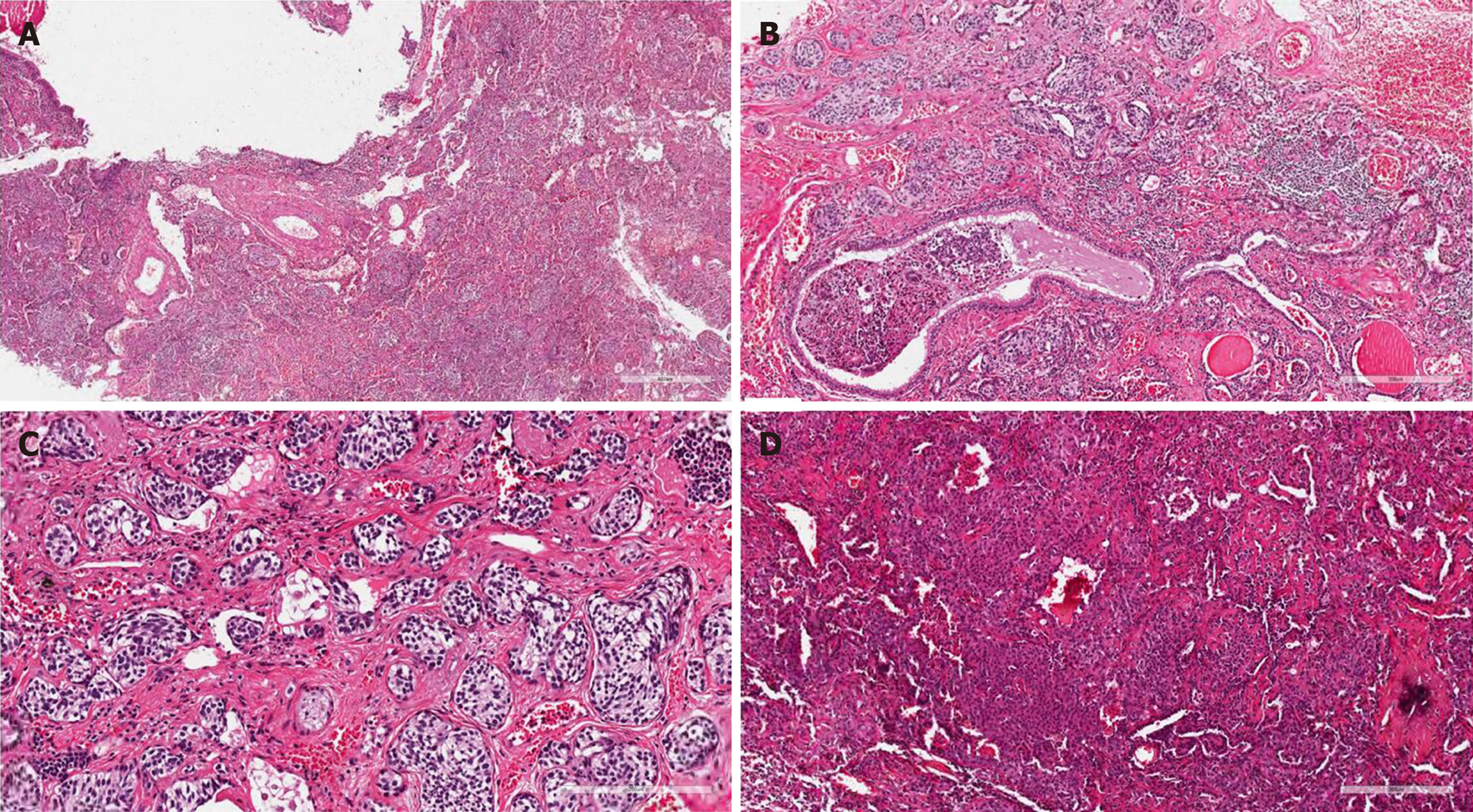
Figure 3 Pathological findings.
A: Microscopic examination showed inflammatory cell infiltration with lymphocytes, neutrophils, and histiocytes in alveoli. Alveolar septa broke and merged, focal bronchiectasis was observed, and cartilage in the bronchus was damaged [(hematoxylin and eosin staining (HE staining), × 40]; B: Multifocal neuroendocrine-like cell nest hyperplasia was found in the surroundings of the expanded bronchus (single focal diameter < 0.5 cm; HE staining, × 100); C: The nuclei of hyperplasia cells are polygonal or short fusiform with a uniform size. The nuclear chromatin was delicate and in fine particles. The nucleoli were unclear; no mitotic figures or necrosis was detected (HE staining, × 200); D: HE staining (× 100) showed the papillary arrangement of tumor cells and a sample of vascular cavity.
- Citation: Han XY, Wang YY, Wei HQ, Yang GZ, Wang J, Jia YZ, Ao WQ. Multifocal neuroendocrine cell hyperplasia accompanied by tumorlet formation and pulmonary sclerosing pneumocytoma: A case report. World J Clin Cases 2020; 8(16): 3583-3590
- URL: https://www.wjgnet.com/2307-8960/full/v8/i16/3583.htm
- DOI: https://dx.doi.org/10.12998/wjcc.v8.i16.3583


