Copyright
©The Author(s) 2020.
World J Clin Cases. Aug 26, 2020; 8(16): 3440-3449
Published online Aug 26, 2020. doi: 10.12998/wjcc.v8.i16.3440
Published online Aug 26, 2020. doi: 10.12998/wjcc.v8.i16.3440
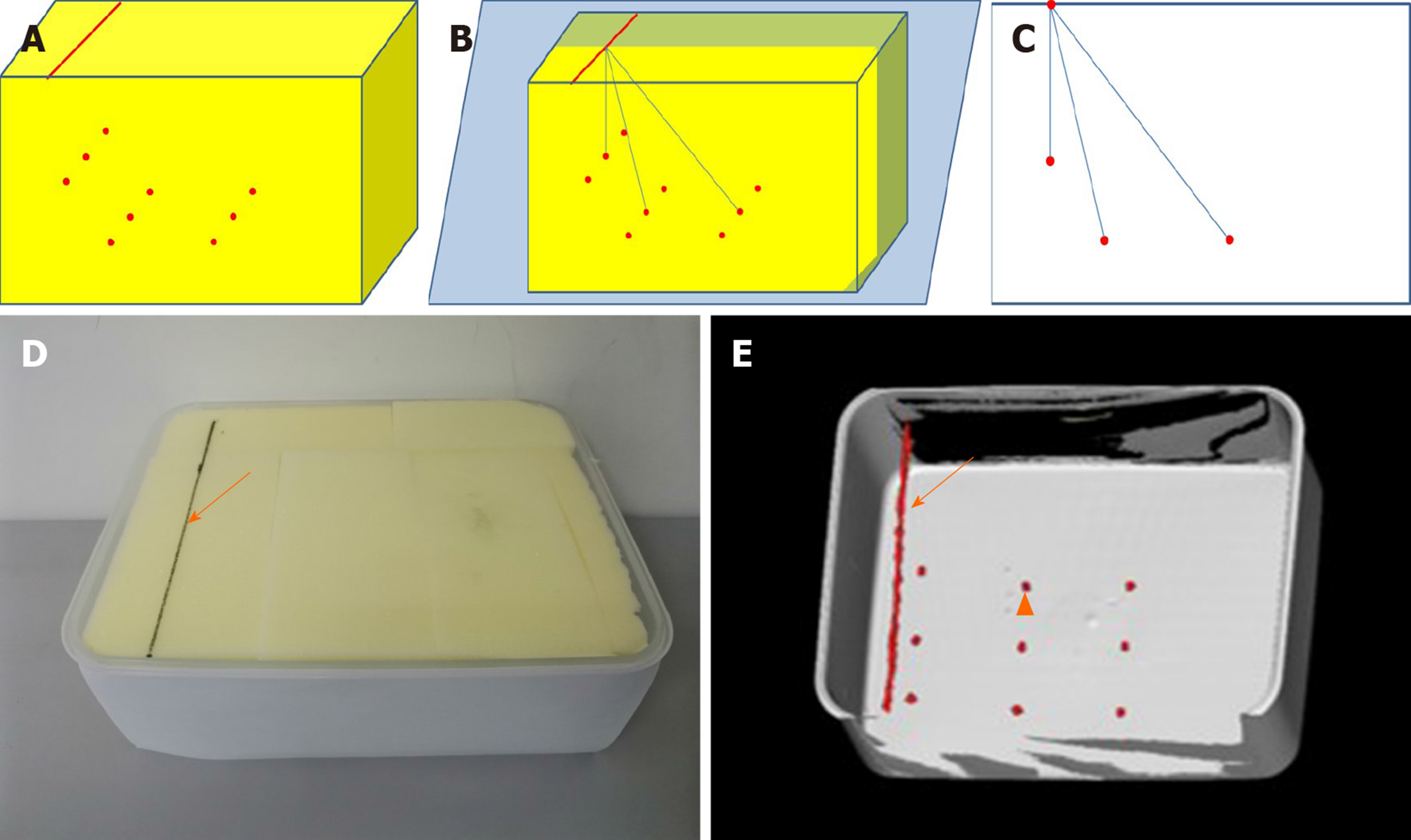
Figure 2 Schematic, photo and three-dimensional computed tomography image of box model.
A: Schematic diagram of box model; B: One insertion line was drawn on the upper surface with contrast agent parallel to the narrow edge; C: Nine artificial targets were set inside the box, respectively 50 mm (target a), 100 mm (target b), 150 mm (target c) far from the insertion line; D: Image and 3D-computed tomography of box model with insertion line and target marked with solid arrow and triangular arrow; E: The box model was made of acrylic resin and filled with sponge.
- Citation: Wang R, Han Y, Luo MZ, Wang NK, Sun WW, Wang SC, Zhang HD, Lu LJ. Accuracy study of a binocular-stereo-vision-based navigation robot for minimally invasive interventional procedures. World J Clin Cases 2020; 8(16): 3440-3449
- URL: https://www.wjgnet.com/2307-8960/full/v8/i16/3440.htm
- DOI: https://dx.doi.org/10.12998/wjcc.v8.i16.3440


